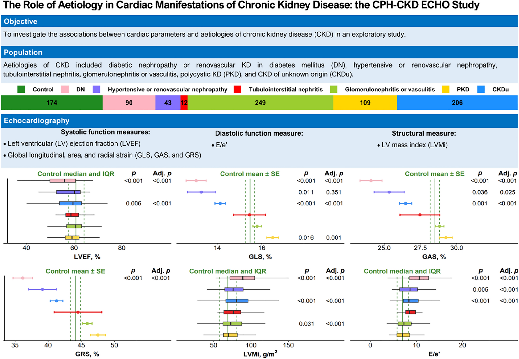
Our objectives were to determine the effect of severe obesity on image quality for cardiac MRI and TTE, and to assess ventricular function agreement between modalities. When compared with patients with normal BMI, image quality for patients with severe obesity remained high and preserved for cardiac MRI but was negatively impacted for TTE. Furthermore, categorical agreement in LV and RV function between cardiac MRI and TTE was worse in the severely obese cohort compared with the normal weight cohort, which may lead to misdiagnosis or inappropriate treatment. These findings highlight the significance of choosing the appropriate imaging modality for clinical assessment and underscore the link between image quality, diagnostic assessment, and subsequent treatment decisions.
This study introduced a novel scoring system to evaluate and compared MRI and TTE image quality in a semi-quantitative manner. The scoring focused on criteria assessable by both modalities, such as ventricular morphology and function, excluding modality-specific criteria like myocardial fibrosis. While prior studies have examined image quality within populations [22] or modalities [23], our approach compared image quality between populations (normal BMI vs. severely obese) and across modalities (MRI vs. TTE). Image quality across all modalities can be compromised in patients with severe obesity [8, 24, 25], and while MRI and TTE are routinely used to evaluate these patients, a head-to-head comparison of the impact of severe obesity on both MRI and TTE had not been previously investigated.
Our finding that TTE image quality significantly degrades in patients with severe obesity is in agreement with previous studies [26, 27]. Increased fat mass causes signal attenuation, reducing the capacity of TTE to assess cardiac morphology, function, and flow [28, 29]. The use of microbubble contrast agent can help to maintain TTE image quality in patients with obesity, although adding to the complexity and cost of the exam [7, 15]. Ellenberger et al. recently reported echo contrast use of 23% of patients with normal BMI and 71% of patients with severe obesity, comparable to the 20% and 64% rates of contrast usage, respectively in our cohorts [30]. We found that patients receiving microbubble contrast in either normal BMI or severely obese cohorts had similar image quality scores as those not receiving contrast. Although contrast may enhance the LV endocardial border, it does not typically improve assessment of valves, vessels, or atria. As supported by Ellenberger et al. and our findings of similar TTE image quality scores with and without contrast, we speculate that microbubble contrast is often used when pre-contrast image quality is severely compromised and does not necessarily improve image quality substantially when pre-contrast quality is poor.
Consistent with the assumption that MRI is the modality least sensitive to image quality degradation caused by obesity [31], we observed no significant difference in cardiac MRI image quality between normal BMI and obese cohorts. Beyond the function and flow measurements afforded by TTE, MRI provides myocardial tissue characterization and the ability to evaluate scar, fibrosis, and edema. In our study, T1 mapping and LGE demonstrated consistent image quality between BMI cohorts, indicating feasibility regardless of body habitus. However, severe obesity poses challenges for MRI as well, including bore size and patient table weight limitations, increased burn risk caused by skin folds, and increased rates of claustrophobia [8]. Lower field, open MRI scanners have been available for some time, offering an alternative that can accommodate patients with severe obesity and/or claustrophobia. While cardiac MRI has been demonstrated on these systems [32, 33], an open magnet configuration can require compromises in overall system performance. The recent introduction of low-field MRI scanners with conventional magnet design but larger bore diameter and higher table weight limits may help expand MRI access to patients with severe obesity [34]. Additionally, low-field systems are significantly less costly to acquire, install, and operate than standard 1.5 T or 3 T scanners, and recent reports are demonstrating good cardiac image quality [35].
When examining differences between cardiac MRI and TTE imaging quality, we found that MRI image quality was superior to TTE in both normal BMI and severely obese cohorts, and this difference was more pronounced in the severely obese cohort. This observed overall image quality effect was also seen in each pre-specified subcategory (chambers, valves, and vessels), suggesting that the overall score difference was not skewed by the effect of a single subcategory. A study conducted by Kanagala et al. also found image quality differences between MRI and TTE in patients with heart failure [22]; the authors attributed this in part to the effect on TTE image quality of common comorbidities including obesity [22, 35].
Cardiac MRI is widely accepted as the reference standard for cardiac morphology and function, and we found discrepancies in MRI and TTE measures of left and right ventricular function with poorer agreement between TTE and MRI in patients with severe obesity compared with those with normal BMI. LVF was misclassified by TTE in approximately one-third of patients with severe obesity, and RVF misclassified in nearly one-half. Notably, RVF by TTE has previously demonstrated varying correlation with MRI [13, 36], and our findings also reflect this real-world variability with no agreement in RVF in patients with severe obesity. Review of the 53 patients with LVF < 50%, only 9% of these had clinical status changes (one arrest, three restoration of sinus rhythm, and one due to medical treatment of heart failure). The remaining LVF discrepancies could not be explained by interval clinical events suggesting the differences in ventricular function may be better attributable to image quality. Despite a majority of patients in both cohorts having normal cardiac function, we demonstrated a significant effect of severe obesity on categorical ventricular function assessment. We speculate that a larger cohort with abnormal cardiac function would have more dramatic observations.
Our study has several limitations. The retrospective design introduces potential referral biases, although no MRI studies were included with an indication of non-diagnostic TTE. We were unable to account for patients not referred to MRI due to claustrophobia or an inability to fit into the scanner bore, and MRI studies that were not completed due to patient discomfort or claustrophobia. Additionally, the retrospective nature of the study and non-contemporaneous scans introduce the possibility of circumstantial events between scans that could impact cardiac function and image quality. Two-thirds of patients had their MRI and TTE performed within 90 days apart, and given that the time differences between TTE and MRI were not statistically different between the normal BMI and severely obese cohorts, we expect that any potential time-related biases would have been equivalent (Supplemental Data). While our novel image quality scoring system was not previously validated, the scores confirmed the quality degradation due to severe obesity by TTE but preserved in cardiac MRI. Future prospective studies employing simultaneous MRI and TTE assessments with standardized LVF and RVF quantification methods would help mitigate any biases introduced by the time between exams.
Our cohorts comprised only normal weight and severely obese patients and did not include those falling into overweight to moderately obese categories (25 < BMI < 40). Therefore, we cannot draw any conclusions regarding MRI or TTE image quality in these intermediate groups. Finally, all MRI scans were done at 1.5 T. Cardiac MRI is also commonly performed at 3 T, which offers higher signal-to-noise ratio that can be used to increase spatial resolution or shorten scan time. However, artifacts caused by field inhomogeneity are worse at higher field, thus it is unclear how our results would translate to 3 T MRI.
In conclusion, cardiac MRI may provide more reliable diagnostic information than TTE in patients with severe obesity, suggesting cardiac MRI could be used as the first-line modality choice for CVD evaluation in this population. Cardiac MRI image quality was preserved in patients with severe obesity, whereas TTE showed a significant quality degradation in this group. Contrast-enhanced TTE did not demonstrate superior image quality compared to non-contrast exams in either patient cohort. Furthermore, there was worse agreement between MRI and TTE in the categorical assessment of LVF and RVF in patients with severe obesity. With a high and increasing prevalence of severe obesity [2], high quality and accurate imaging techniques are necessary to support best clinical practice.


留言 (0)