
記住我


A 53 year-old male patient presented at the emergency department with unstable angina pectoris since 24 h. He experienced similar symptoms a week before presentation when he had a cough and signs of pneumonia successfully treated with 600 mg Ibuprofen 3-times daily. He is smoker and his father died at the age of 53 due to acute myocardial infarction. Upon admission, the patient is febrile (38.5 °C) with respiratory frequency of 12/min, peripheral O2 saturation 96% under room air, sinus rhythm (77 beats/min) and blood pressure of 128/79 mmHg. The pH is 7.341, arterial lactate 0.4 mmol/L and venous lactate 1.6 mmol/L. High-sensitivity cardiac troponin I is 3600 ng/L and CK-MB is 40 U/L.
Coronary angiography revealed an abrupt occlusion of the first marginal branch of the circumflex artery (Fig. 1A) without collateralization and an otherwise intact left dominant coronary artery system (Fig. 1B). Ventriculography raised the suspicion of a small left ventricular pseudoaneurysm (Fig. 1C). Transthoracic echocardiography revealed normal left ventricular function and volumes, left lateral wall dyskinesia and a small pericardial effusion. The CT angiography (Fig. 2A) confirmed the suspicion of small anterolateral left ventricular pseudo-aneurysm (red circle) without sign of free rupture and a minor pericardial effusion (red arrow).
Fig. 1
Preoperative screening for coronary disease in patient I Preoperative coronary angiography of patient I reveals occlusion of the first marginal branch (red arrow) with retrograde perfusion from the posterior interventricular branch (black arrow) of the left dominant coronary system (Fig. 1A), intact small right coronary system (Fig. 1B) and the suspicion of left ventricular pseudoaneuryms (red circle) without pericardial effusion of contrast substance (red arrow) in ventriculography (Fig. 1C)
Fig. 2
Preoperative thoracic CT-Angiogram Fig. 2A shows minimal pericardial effusion (red arrow) with suspicion of LVFWR of the first marginal branch (red circle) in patient I Fig. 2B shows relevant pericardial effusion (red arrow) and a hypocontrasted myocardial area of the first marginal branch (red circle) in patient II
The patient remained hemodynamically stable and clinically asymptomatic during and after the examinations and was transferred to the ICU for continuous surveillance. High-sensitivity Troponin I decreases from 3600 ng/L to 2300ng/L and CK-MB from 40 U/L to 26 U/L. Pro-BNP increases from 693 pg/mL to 1156 pg/mL (Table 1). The patient remained awake, asymptomatic, catecholamine-free and pulmonary stable with 2 L nasal oxygen. Cardiac MRI was planned for the next morning. Twenty hours after hospital admission, hemodynamic instability occurred followed by instant circulatory collapse. Cardiopulmonary resuscitation was initiated with intubation and mechanical ventilation. Ventricular fibrillation could not be converted to sinus rhythm. Echocardiography revealed relevant pericardial effusion. Pericardial drainage was performed and more than 1 L fresh arterial blood removed. Persistent electromechanical dissociation was assessed. Veno-arterial ECMO was placed through percutaneous femoral approach under massive volume resuscitation (2500 mL crystalloid). High-dose cathecolamine support (120 µg/min Noradrenalin) was required to maintain circulation. The patient was immediately transferred to the operation room (OR), that was 21 h after the hospital admission, with arterial Hb 5, pH 7.2 and lactate 20.9. Sternotomy was performed under ECMO support. Bradycardic broad QRS complexes and cardiac systolic ejection appeared after opening the pericardium and removing the massive pericardial clot (> 1000 ml). After full heparinisation, the peripheral ECMO cannulas were connected to the heart-lung machine and 6 units of packed red blood cells (PRBC) were administered immediately.
Table 1 Perioperative and operative patient dataInspection of the unloaded heart on cardiopulmonary bypass (CPB) revealed a 2.5 cm long LVFWR within a 1 × 3.5 cm necrotic area in the region of distribution of the first obtuse branch of the circumflex artery (coronary segment 12) confirming with the previous angiography (Fig. 1). Coronary calcification was excluded by inspection and palpation. Epicardial echography showed significantly reduced global biventricular function with akinesia of the lateral wall of the left ventricle and ruled out valvular abnormalities.
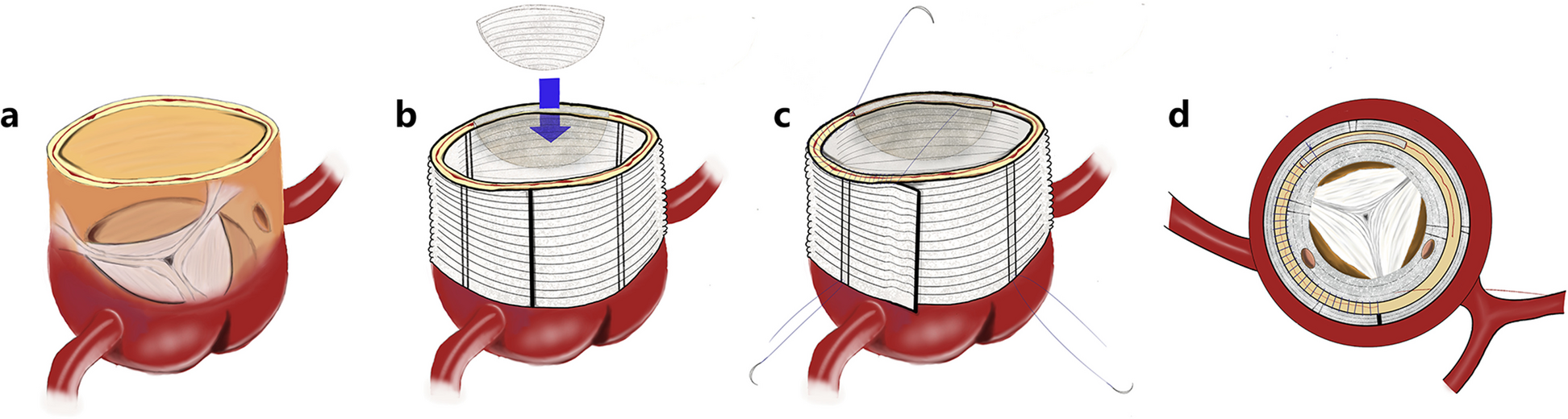
Aortic cross-clamping and cardioplegic cardiac arrest was instituted by intermittent antegrade and retrograde administration of blood warm (Calafiore) cardioplegia. Surgical repair of the left ventricular tear was performed by linear closure with Prolene 4/0 as a horizontal mattress sutures with two supporting 4 cm long Teflon-felt strips. A second patch of Teflon was sealed on the suture line using surgical glue and fixed with an additional running Prolene 4/0 suture on the mattress suture. A hemostatic TachoSil (hemostatic fleece) was applied to widely cover the suture and the adjacent left ventricular tissue. Controlled aortic root reperfusion was instituted for 10 min. No electrical activity was assessed. Aortic declamping followed. Reperfusion was continued under epicardial DDD-pacing and lung-ventilation. Further transfusion and adequate ultrafiltration were performed (Table 2). Although trasesophageal echocardiography ruled out valvular dysfunction or regional myocardial dyskinesia, weaning from the CPB was not possible due to persistent biventricular cardiac failure. The ECG showed a bradycardic broad QRS ventricular rhythm hardly reacting to DDD-pacing. After 60 min of reperfusion, CPB circulation was switched back to veno-arterial ECMO support. This maneuver was not well tolerated by the patient, and shortly thereafter cardiac arrest was assessed in echography. Although massive inotropic and chronotropic support with Dobutamin, Milrinon, Noradrenalin, Vasopressin and finally Adrenalin was applied, stable hemodynamics could not be achieved. Venous return decreased progressively and continuous volume substitution was necessary to maintain ECMO flow. Intraabdominal free fluid accumulation was not detected by sonography. No significant intrathoracic bleeding was observed. Lactate values remained high in spite of the extensive therapy (Table). Massive volume administration failed to improve circulation and the patient died 50 min later under maximal pharmacological support with ECMO-flow decreasing below 1 L/min, blood pressures below 50 mmHg and lack of electrical response to maximal epicardial pacing.
Table 2 Intraoperative patient dataPatient IIA 76 year-old male patient without previous medical history and no known cardiovascular disease presented to the emergency department of a district hospital due to sudden syncope and progressive dyspnea. Upon admission the patient shows arterial hypotension (96/53 mmHg), tachycardia (88beats/min), jugular venous congestion, respiratory frequency of 18/min, peripheral O2 saturation 90% under mask oxygenation with 10 L 100% O2. He is somnolent, acidotic (pH 7.184) with elevated lactate (arterial 8.4 mmol/L, venous 9.6 mmol/L).
Transthoracic point-of-care echocardiogram showed relevant circumferential pericardial effusion (> 2 cm) with compression of the right atrium and ventricle. The patient was emergently transferred to our university department with the suspicion of Type A acute aortic dissection under increasing continuous catecholamine administration (achieving 40 µg/min noradrenaline). The ECG showed no signs of acute myocardial infarction. The emergent CT-Angiography (Fig. 2B) revealed normal ascending and descending thoracic aorta with no signs of dissection, a hemodynamically relevant (> 700 ml) pericardial effusion (red arrow) and a hypocontrasted myocardial area (3 × 2 × 2 cm) on the lateral left ventricular wall (red circle). Three-dimensional reconstruction of the coronary CT-angiography revealed normal coronary ostia with minimal calcification of the main branches (Fig. 3A, B and C). High-sensitivity cardiac troponin I was 3006.2ng/L and CK-MB < 24U/L.
Fig. 3
Preoperative screening for coronary disease in patient II Coronary thoracic CT-angiogram reconstruction of patient II reveals an intact main left coronary artery (Fig. 3A), suspicion (red arrow) of an occluded first marginal branch (Fig. 3B), and an intact right dominat coronary artery (Fig. 4C)
The patient was increasingly unstable during the examinations, with progression of hypotonia and tachycardia in spite of the higher catecholamine substitution (80 µg/min Noradrenaline). Under the diagnosis of critical pericardial tamponade emergency operation was indicated and the patient was immediately transferred to the OR without previous coronary angiogram, and arrived in the OR 2 h after his presentation to the district hospital with laboratory revealing Hb 14, pH 7.2 and lactate 9.8. The patient remained awake and breathing independently with oxygen mask.
Insertion of the central venous and peripheral arterial lines, disinfection, draping and preparing the operating table for CPB took place before initiation of anesthesia. Asystole occurred during relaxation and endotracheal intubation. Sternotomy was performed under intermittent mechanic resuscitation in less than 2 min from the time of endotracheal intubation. Spontaneous circulation recovered immediately after pericardiotomy and removal of clot (> 800 ml). Sinus rhythm and elevated blood pressure occurred after 1000 mL crystalloid volume administration. Inotropic support was paused. Inspection of the heart revealed a 2.0 cm long LVFWR within a 1 × 3.5 cm necrotic area in the region of distribution of the first obtuse branch of the circumflex artery (coronary segment 12) and conforming with the hypoperfused territory detected on the CT scan (Fig. 2B). Inspection and palpation revealed a right dominant coronary system with no signs of coronary calcification. Transesophageal echocardiography showed good biventricular function, normal coronary ostia, normal ventricular wall motion and ruled out valvular pathology. Cannulation of the ascending aorta and double stage right atrial cannulation were performed and CPB instituted.
Aortic cross-clamping and cardioplegic cardiac arrest was instituted by intermittent antegrade and retrograde administration of blood warm (Calafiore) cardioplegia. Surgical repair of the LVFWR was performed by linear closure with a Prolene 4/0 horizontal mattress sutures with two supporting 3.5 cm long Teflon-felt strips. Surgical glue was applied along the suture line and a TachoSil fleece was applied to cover the suture and the additional left ventricular tissue.
Controlled aortic root reperfusion was instituted for 5 min and a normo-frequent sinus rhythm was assessed. Aortic declamping and transition from mechanical pump-assisted circulation to spontaneous heart activity was easily achieved with sufficient blood flow to maintain systemic circulation, under minimal catecholamine support. Weaning from CPB was performed under echographic surveillance. Good biventricular function without wall dyskinesia was assessed. Valve function was normal. Three units of FFP and 2 gr Fibrinogen were administered after ending CPB (Table 2). Lactate fell below 4 mmol/L (Table 1). Decannulation, hemostasis, sternal and wound closure were performed. The patient remained stable and was transferred to the ICU under minimal catecholamine support. Extubation followed 6 h postoperatively. Neurological examination was flawless. Oral nutrition and mobilization were initiated. The patient was transferred to the ward on the second postoperative day and to the rehabilitation on the 7th postoperative day. Coronary CT-scan performed before hospital discharge showed minimal and localized coronary calcifications without relevant stenosis. Full clinical recovery was achieved. Twelve months postoperatively, the patient is living at home symptom-free, in sinus rhythm, with normal biventricular pump function on aspirin, statin and minimal beta-blocker therapy.
留言 (0)