
記住我


Surgical treatment of infiltration or thrombosis of the inferior vena cava (IVC) in malignant liver neoplasms is associated with high risks, which are counterbalanced by possible advantages in the absence of alternative therapies [1]. In the absence of surgical treatments, the median survival rate is less than 12 months, and chemotherapy has demonstrated acceptable survival rates in few reports [2, 3]. In this report, we demonstrate the surgical treatment of a patient with a liver metastasis complicated by the thrombosis of IVC and right atrium undergoing cardiopulmonary bypass (CPB).
This work has been reported in line with the SCARE 2020 criteria [4].
Case presentationA 60-year-old woman with a clinical picture of dyspnea at moderate physical exertion, weakness and discomfort in the heart area was admitted to the Clinic of Faculty of Surgery No. 1. In 2017, a left-sided hemicolectomy was performed for splenic flexure transverse colon cancer pT4AN2AM0 (TNM 8), histologically moderately differentiated adenocarcinoma. The patient received a total of 8 courses of adjuvant chemotherapy on the CAPOX scheme in 2017. The patient was further operated, twice for liver metastasis: in 2018, liver segments 2, 3 and 4 A were resected; in 2019, liver segmentectomy IVB was performed. After liver segmentectomy IVB patient received 8 courses of adjuvant chemotherapy on the FOLFIRI scheme in 2019 with dose reductions to avoid intolerance. We performed an echocardiography on January 2020 that showed a subtotal thrombosis of the right atrium, and other parameters were normal. Computed tomography (CT scan) revealed a hepatic segment I metastasis with inferior vena cava thrombosis, spreading into the right atrial cavity (Fig. 1 A, B).
Moving thrombotic masses in the right atrium were confirmed by transesophageal echocardiography (TEE). (Fig. 1 C).
Surgery was performed on February 5, 2020: median sternotomy and upper median laparotomy. The stage of mobilization and removal of the first segment of the liver was performed before the start of CPB. Cardiopulmonary bypass was connected by type: ascending aorta, superior vena cava and inferior vena cava (below tumor thrombosis) under normothermic conditions. After adhesiolysis, tourniquets on the superior vena cava and IVC were placed. Tourniquets are superimposed together to place venous cannulas and are squeezed so that blood does not fall below the place of standing of the cannula of the superior vena cava and above the place of standing of the cannula of the inferior vena cava. Longitudinal cavatomy was performed, the revision revealed invasion of the inferior vena cava wall up to the entry into the right atrium.
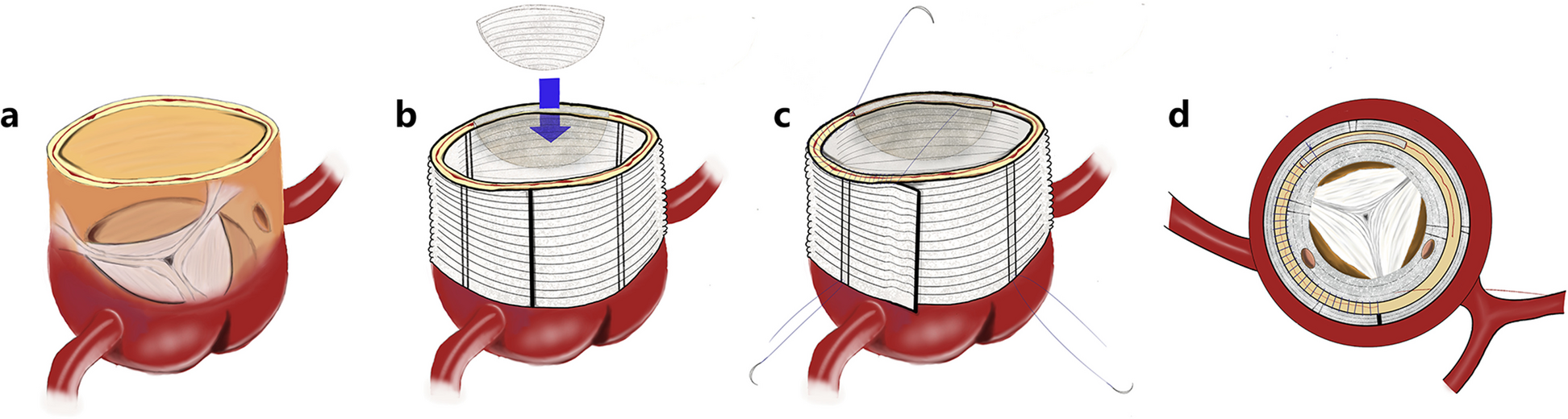
Thrombotic masses were visualized, which spread into the right atrium and were fixed to the wall, the tricuspid valve was intact. Histological examination revealed tubular adenocarcinoma of the liver and R0 edge of resection. For radical removal of the tumor conglomerate, to reduce the time of CPB and more physiologically close the major defect of the right atrium and prosthetics of the inferior vena cava, we decided to implant a pulmonary cryopreserved homograft in the position of the inferior vena cava. We performed resection of the area of the right atrium and inferior vena cava involved in the tumor process, and prosthetics of the area of inferior vena cava, right atrium with cryopreserved pulmonary homograft of 28 mm in diameter. The proximal anastomosis was formed with the widest part of the pulmonary homograph (bifurcation area), the distal anastomosis was formed with the proximal portion of the pulmonary homograft, the pulmonary valve was previously excised. The proximal and distal anastomoses were formed by a continuous winding suture using a prolene 5 − 0. The cardiopulmonary bypass time was 67 min.
Tumor, a part of the resected inferior vena cava with part of right atrium, pulmonary homograft are showed after reconstruction in (Fig. 1 D, E,F).
After surgery, we observed the patient in the intensive care unit for 24 h, and then the patient was transferred to the cardiac surgery unit, where she continued her rehabilitation. On the first day we removed postoperative drains from the thoracic cavity. On the 3rd day we removed the drainage from the abdominal cavity as well. The postoperative period was smooth and the postoperative wounds healed. However, in the postoperative period (2–3 days) due to the large wounded surface the patient began to feel dyspnea and decrease in saturation up to 90–91%. On the background of CPAP therapy for 5 days the patient’s condition showed positive dynamics, saturation 95–97%. The patient received low-molecular-weight heparin in therapeutic dosage for the first 3–4 days. Then the patient was prescribed rivaroxaban 20 mg continuously.
In postoperative period patient received 4 courses of adjuvant chemotherapy on the FOLFIRI scheme in 2020.
Fig. 1
CT scan before operation, tumor trombosis into right atrium and inferior vena cava (A,B), TEE view before operation demonstrate tumor mass into right atrium (C), resected part of IVC and right atrium (D), cryopreserved pulmonary homograft 28 mm (E), final view (F)
Three year after the operation according to echocardiography and CT, there was no evidence of recurrence (Fig. 2), and an adequate function of the tricuspid valve.
Fig. 2
CT scan after 3 years. There is no data for recurrent
留言 (0)