
記住我

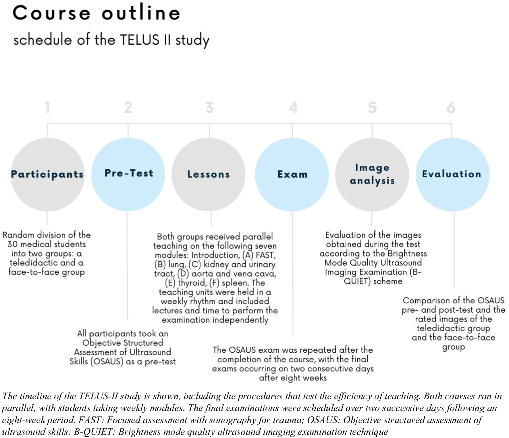


This study is a follow-up project and was supervised by two German Society for Ultrasound in Medicine (DEGUM) certified physicians (level I and level III). In the past, the course was already conducted as a proof of concept study using an online-only format (the TELUS I trial),16 as due to COVID restrictions, no hands-on teaching could take place with a control group. The aim of this study is to compare the learning success of teledidactic teaching with that of the face-to-face group that is now being provided. Participating students were in the clinical part of their medical studies and they could get credit for the ultrasound course as an elective subject. Within their degree program, students must select elective courses, allowing them the freedom to choose subjects based on their interests. The participating students have independently opted for this specific elective. Subsequently, they were randomly assigned to the teledidactic group and the face-to-face group (Fig. 1).
Figure 1
Timeline of the TELUS II study.
Each participating student received a mobile ButterflyIQ17 probe (Version 2, Butterfly Network Inc, Delaware, USA) for the duration of the course and, if necessary, an Apple iPad (Generation 9, Apple, Cupertino, USA) with the corresponding app to operate the probe.
Course OutlineThe course consisted of seven modules:
(0) Introduction to ultrasound
(A) Focused assessment with sonography for trauma (FAST)
(B) Lung
(C) Kidney and urinary tract
(D) Aorta and vena cava
(E) Thyroid gland
(F) Spleen
The selection of the modules was based on the recommendations of an international consensus conference for undergraduate medical students.18 An introductory lecture was given at the beginning of each lesson. In the lecture, the guidance of the ultrasound probe during the examination was outlined. In addition, ultrasound images of typical pathologies were shown. In this way, the clinical reference was not missing, as the students examined each other or friends who were mostly young and healthy. The same slides were used online and face-to-face. After explaining the theoretical knowledge and guidance of the ultrasound probe, the teachers demonstrated the examination. During the online lessons, the camera image and the iPad screen of the teacher were shared simultaneously so that the students could follow the handling of the ultrasound probe (Fig. 2).
Figure 2
Demonstration of the implementation of the teledidactic ultrasound course.
In the face-to-face course, the lecturer demonstrated the examination live with the help of a volunteer, who served as the model. After pending questions were clarified, the students had time to perform the examination on their own. In the online course, they had to organize a volunteer at home to perform the ultrasound examination, while the students assigned to the face-to-face course examined each other. During the lecture, the components that the students were required to include were thoroughly explained to them. For instance, they were told to take a longitudinal and cross-sectional slice of the kidney and measure its length, width, and thickness. While the students performed the examination on their own, the tutors provided direction and support. There were three tutors available for questions and corrections, so that approximately five students were supervised by one tutor. The instructor was able to correct students’ use of the ultrasound probe in the face-to-face course, as opposed to the online course, where assistance was only given in the form of words and videos. The captured ultrasound images were uploaded to the cloud using the Butterfly app. For the students from the teledidactic course to receive direct feedback on their images, the comment function of the Butterfly app was used and the teacher annotated the images to indicate how the presentation of the organs could be optimized. Direct feedback on images uploaded to the cloud was provided during the course upon student requests for clarification or when they faced challenges in recognizing organ structures. Furthermore, comprehensive feedback on these images, including specific comments, was delivered within the same week, ensuring timely responses prior to the subsequent module.
Assessment InstrumentsA suitable assessment tool had to be chosen to evaluate the students’ learning progress and compare the two groups. To test the various competencies, a combination of a practical test and an image rating was chosen. The Objective Structured Assessment of Ultrasound Skills (OSAUS)19 is an assessment tool explicitly designed for ultrasound examinations. The examination protocol can be used for the scanning of different organs and does not need to be specially adapted. OSAUS contains seven fields of evaluation, namely: indication for the examination, applied knowledge of ultrasound equipment, image optimization, systematic examination, interpretation of images, documentation of examination, and medical decision-making. Since a young, healthy patient was selected as the examination model, the scale had to be adapted. For instance, in the field medical decision-making of the thyroid gland, the keyword thyroid inferno was mentioned and asked in which disease this would occur, or in the vena cava examination, reasons for congestion of the vein had to be listed. Before the course, an OSAUS pre-test was conducted with the students to determine their level of knowledge and after completion of the course, the OSAUS exam was repeated to have a direct comparison. In the pre-test, not all modules were covered, but the protocol was limited to the FAST examination, the kidney, urinary tract, and thyroid gland. On the final exam, all the modules that were taught were tested, with the examination of the spleen integrated into the FAST examination, as the recessus splenorenalis had to be visualized. Various fabricated patient case scenarios served as the exam’s guides so that a clinical environment could be simulated. The students had a total of 25 min for each exam, giving them 5 min for each task to demonstrate the inspection of the corresponding organ. During the exam, every student, whether in a face-to-face or teledidactic group, scanned the same person. As a result, the test circumstances were consistent, making it impossible for the person’s prior practice with the test to have an impact. Using the brightness mode quality ultrasound imaging examination (B-QUIET) scale, ultrasound pictures from the final exam were evaluated.20 B-QUIET is a scale for ultrasound image evaluation and contains, among others, the item’s gain, depth, and resolution (Fig. 3). Three independent evaluators, consisting of two physicians and a peer tutor, conducted the lessons, assessed the images, and administered the pre- and post-OSAUS examination.
Figure 3
Evaluation criteria of the brightness mode quality ultrasound imaging examination technique (B-QUIET).
The OSAUS and image rating results of the face-to-face group were compared with those of the teledidactic group to determine whether online teaching can compete with the traditional way of teaching.
Statistical AnalysisThe statistical analysis was realized with RStudio (version 2022.07.1 + 554) and IBM SPSS Statistics 28. Means and standard deviations were calculated as descriptive parameters. Differences were found to be statistically significant when p < 0.05. A Levene’s test was performed to test for equality of variance followed by an independent t-test or single-factor ANOVA to test the null hypothesis indicating that there is no difference between the two groups. Using the B-QUIET scheme, a total of 450 images that were generated in the examination were rated by three independent raters, who also conducted the final OSAUS exam with the students.
The local ethics committee of the university approved the study and all enrolled students gave written informed consent to the participation in the course and to the use of their images. For managing incidental findings, we have utilized an article that provides a framework for defining and handling them within the context of ultrasound courses.21
留言 (0)