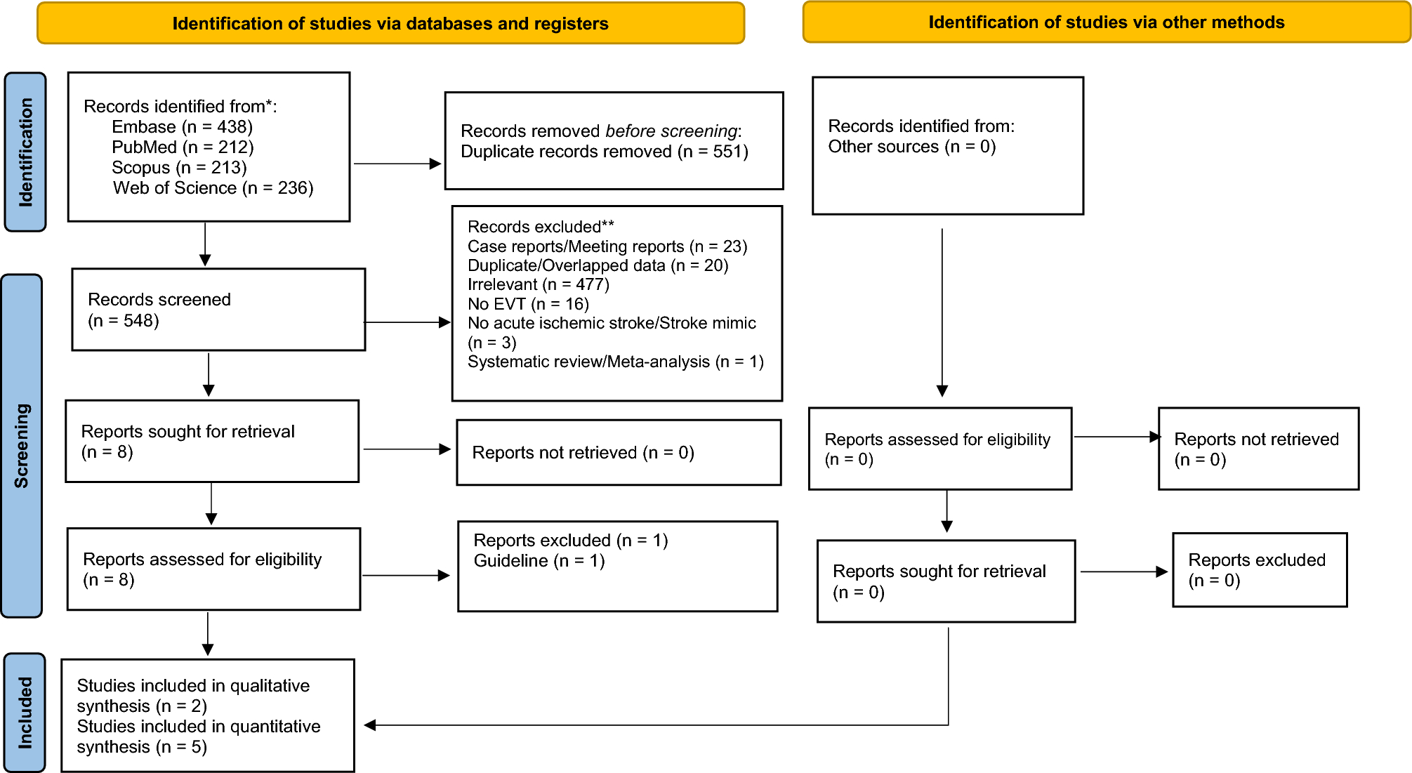
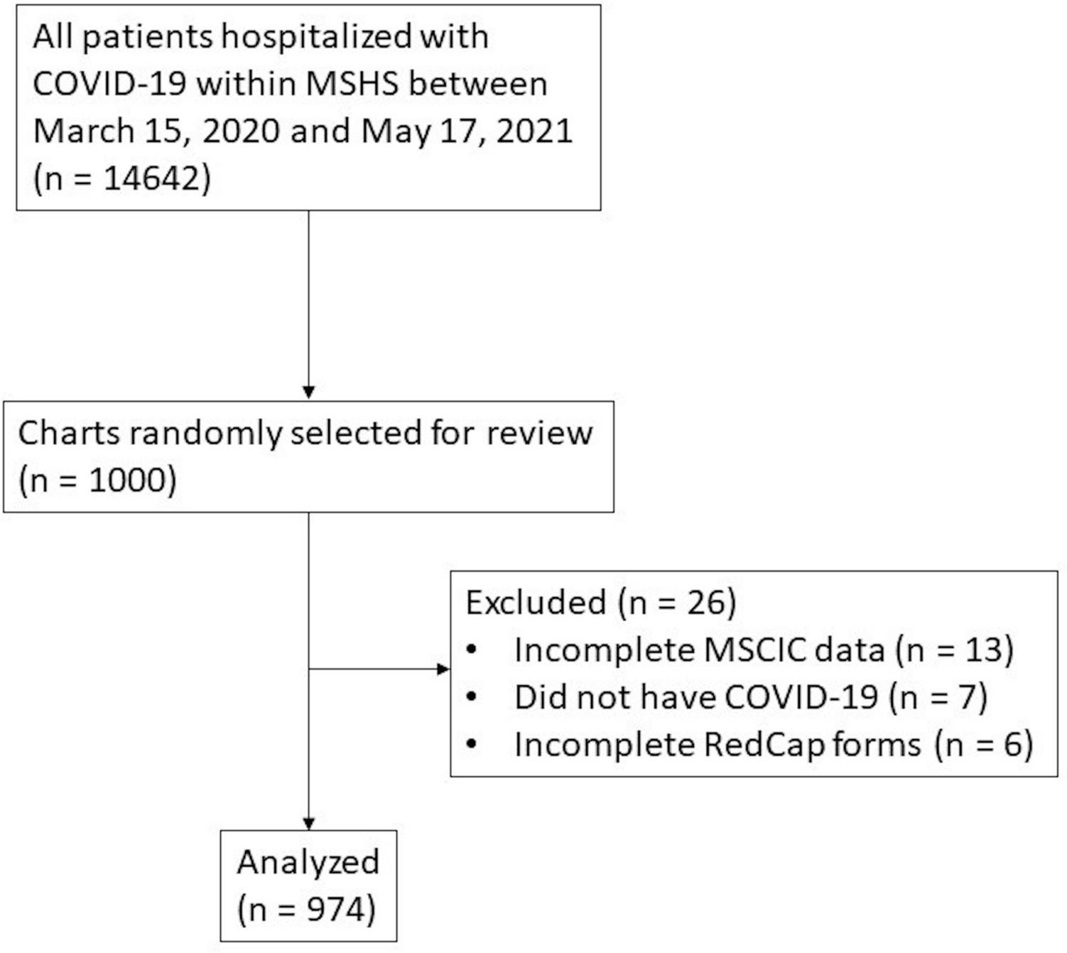
RT-QuIC testing, with its extremely high specificity, has transformed the diagnosis of sporadic CJD and has allowed for more accurate in-life diagnosis. Results from our study demonstrate that a small yet significant portion (~ 10%) of those with sporadic CJD have a false-negative RT-QuIC result when diagnosis is confirmed at autopsy. It is important for clinicians to understand the clinical characteristics and performance of other investigation results in this patient population. Furthermore, this study aimed to explore the contribution of underlying molecular subtypes on performance of RT-QuIC in this population and confounders relating to co-pathologies identified at autopsy.
This study identified that individuals with a negative RT-QuIC were likely to be younger at diagnosis and have a longer disease duration compared to those with a positive RT-QuIC. This finding confirms previous reports [8,9,10]. Of these three previous studies, only Jones et al. specifically noted the median age (RT-QuIC negative, 52; RT-QuIC positive, 66) and duration of disease (RT-QuIC negative, 710 days (~ 23.7 months); RT-QuIC positive, 147.5 days (~ 4.9 months)). Our study identifies a more modest difference between the median age of onset (RT-QuIC negative, 62 years; RT-QuIC positive, 68 years) and duration of disease (RT-QuIC negative, 10.5 months; RT-QuIC positive, 4.4 months). A reason for this variation may be related to smaller sample size (n = 13) and study design as Jones et al. included both those with definite and probable sCJD.
Our study also found those with a negative RT-QuIC were more likely to note sleeping difficulties as an initial symptom and at initial diagnosis were less likely to have motor symptoms and difficulties with gait. Foutz et al., in their study on the sensitivity/specificity of RT-QuIC, reported on symptoms in those with a negative RT-QuIC identifying that that they were significantly less likely to present with ataxia [9]. Jones et al. also identified that in those with RT-QuIC negative ataxia was less frequent (31%) compared to positive patients (61%), however, this difference was not found to be significant (p = 0.07) [10]. RT-QuIC negative individuals in our cohort were less likely to have difficulties with gait (73% vs 93%) or present with motor symptoms (62% vs 80%) at diagnosis. However, due to performing multiple tests of comparisons our result regarding reduced motor symptoms (p = 0.04) should be interpreted with caution as it only just exceeds our threshold for significance.
Demographics and clinical features of RT-QuIC individuals are likely explained to some extent by the overrepresentation of certain underlying molecular subtypes and reflective of the characteristics of their individual clinico-pathological phenotype [11]. Those with the VV1 subtype of sporadic CJD have a younger age of onset (39–44 years) with a prolonged disease duration (15–21 months) [11, 15]. Although in depth clinical phenotyping of this sub-group is challenging due to its rarity (~ 1% of those with sporadic CJD), these individuals most commonly present with a slowly progressive dementia with psychiatric changes with a lower degree of neurological signs and symptoms on first examination [11, 15]. The low presence of gait abnormalities/motor symptoms when first diagnosed may simply reflect the earlier stage they are in their disease process. Our findings are comparable to these studies identifying that in our cohort of RT-QuIC negative individuals with definite CJD that those with the VV1 subtype had a median duration of 17.28 with a median age of onset of 53. The MM2C subtype also presents with a longer disease duration as a progressive dementia with a slower progression of neurological symptoms. Interestingly whilst our study also identified that those with the MM2 had a longer median duration of 30.24 months they also presented with a younger age of onset of 57 years which is not typical of this subtype [16, 17]. This finding is likely explained by our small sample size (n = 4) and larger studies are needed to assess its significance. Overall, it is likely that the presence of these subtypes and their associated clinical features explain why those with a negative RT-QuIC are more likely to present with a younger age of onset and longer duration of disease.
In our cohort, individuals with negative RT-QuIC were more likely to describe sleep difficulties as an initial symptom compared to those with a positive RT-QuIC. Of these four, only two had molecular subtyping (MM2 and MV1) with the other two only possessing codon 129 genotyping (VV and MV). None of our cohort had the MM2T subtype of CJD (sporadic fatal insomnia). Interestingly two of the four with sleeping difficulties also had significant co-pathology in the form of severe amyloid beta deposition (both Thal V) and neurofibrillary tangles corresponding to Braak & Braak IV and V. However, due to the small sample size (n = 4) it is difficult to draw conclusions regarding its significance.
Performance of CSF 14-3-3, DW-MRI, and EEG
Our study is in line with previous studies that demonstrate that there is no significant difference between the sensitivity of MRI or EEG in those who are RT-QuIC negative and positive but that 14-3-3 demonstrates a lower sensitivity in RT-QuIC negative cases [9, 10]. The sensitivity of MRI was found to be high in those with definite CJD with a negative RT-QuIC (96%) with no significant difference found when comparing with those who were RT-QuIC positive. There was no difference demonstrated between the areas of diffusion restriction on DW-MRI between those who were RT-QuIC positive or negative. Due to the overrepresentation of these two subtypes (MM2 and VV1) it may be expected that this cohort would be more likely to have cortical ribboning on imaging. This is because several case series have identified that in both of these subtypes they are more likely to present with cortical ribboning with less frequent involvement of the basal ganglia [10, 15,16,17,18]. Our study findings indicate that MRI is highly sensitive in sCJD irrespective of RT-QuIC outcome. This should support clinicians tasked with making difficult decisions around palliative management in sCJD as well as public health-related actions.
EEG was found to have a low sensitivity (30%) which points to the lack of clinical utility of this test in aiding the diagnosis of sCJD in those who are RT-QuIC negative. This is slightly higher than a previous study comparing RT-QuIC negative and positive cases (16%) [10]. Furthermore, the VV1 and MM2C subtype are associated with lower sensitivity of EEG and clinicians would be advised to interpret a non-diagnostic result with caution [15, 16]. One hypothesis may be that this is due to the longer duration associated with these subtypes and this represents EEG likely finding less utility in those earlier in the disease process. Another is that these subtypes themselves are simply less likely to show periodic sharp wave complexes and further studies should attempt to explore these two possible hypotheses.
Our study identified that 14-3-3 has reduced sensitivity in those who are RT-QuIC negative, in concordance with findings of previous studies [8, 9]. This is likely secondary to a combination of reasons. The 14-3-3 protein family is highly expressed in the brain and, while raised in CJD, are a non-specific marker of neuronal damage and can be raised in strokes, seizures, paraneoplastic syndromes, and autoimmune encephalitis [19, 20]. As 14-3-3 is a marker of neuronal damage, it has been shown to have increased sensitivity the later in the disease course that the CSF is sampled [21]. Furthermore, 14-3-3 has been demonstrated to have reduced sensitivity in the MM2C and VV1 subtypes and this is another possible explanation for these results [11, 13, 14]. This is supported by our findings that only 0/4 of those with MM2C and 2/6 of those with VV1 had a positive 14-3-3.
Molecular subtype and neuropathology
Results from our study are in line with those of previous studies identifying that those with the MM2 and VV1 subtypes are over-represented in those with a negative RT-QuIC [1, 8, 9]. It is currently unclear why these molecular subtypes are unlikely to test positive on RT-QuIC. It is unlikely that longer disease duration, and possible reduced protein seeding from earlier sampling, are the cause as studies have identified that RT-QuIC is unaffected by timing of CSF collection [14]. Furthermore, RT-QuIC can even be positive in individuals with prion disease prior to symptom onset [22]. A more likely answer is that some underlying characteristic of the prion protein in those with the VV1 and MM2 subtypes causes reduced protein seeding and thus reduced fluorescence on RT-QuIC testing. Studies evaluating the effect of different CJD subtypes on RT-QuIC positivity have identified an increased lag time on VV1 testing and in the study by Peden et al. increased lag time on VV1 and MM2C testing [23, 24]. A study by Poleggi et al. reviewing the effect of different recombinant prion protein substrates on VV1 and MM2C identified similar findings. Interestingly they also found that there was increased sensitivity in the improved QuIC (IQ-CSF) compared to the prior protocol QuIC (PQ-CSF) in those with MM2C (70% vs 40%) but this was found not to be the case in those with VV1 [25, 26]. The reduced PrPsc seeding efficiency in these subtypes may correlate with the atypically long durations when compared to the other variants of sporadic CJD with a median duration of 3–5 months.
Strengths and weaknesses of the study
The UK NCJDRSU surveillance cohort, as a centre for national referrals, has a high rate of case ascertainment as well as dedicated clinical phenotyping via structured clinical assessments (completed by a trained clinician). This study is also the first to directly compare the clinical, imaging, genetic, and biochemical characteristics in RT-QuIC negative and positive individuals in those with definite CJD. By doing this we aim to help clinicians when interpreting a negative RT-QuIC result and we help further characterise the clinical, imaging, and genetic profile of this cohort. All RT-QuIC test results used in this study have been performed at a single lab with a structured methodology. All imaging interpretation was performed by a neuroradiologist with expertise in prion imaging (D.S.) who is blinded to clinical parameters and reviewed in a standardised format to improve reliability and repeatability. Imaging interpretation was reported by a single individual reducing any variation secondary to inter-rater reliability.
There are some limitations to our study. We identified that those with a negative RT-QuIC and a post-mortem were likely to be younger. Furthermore, while unable to be assessed, it is likely that those with a post-mortem were more likely to have atypical disease progression/symptom profile resulting in an over-representation of those with rare molecular subtypes and those with neurodegenerative co-pathology. However, this is a broader issue affecting any study performed on a post-mortem cohort and there is no reason to suspect that our cohort is different than other studies performed on this population. There was incomplete genetic testing performed (37% of RT-QuIC negative individuals missing data on mutations) which raises the possibility that these individuals may have had genetic CJD. While we noted a high proportion of co-occurring neurodegenerative pathology in those who are RT-QuIC negative we have not compared this to a control group. Further studies should aim to assess if this significantly higher by making a comparison to those with a positive RT-QuIC result. Furthermore, our study utilized multiple tests of comparison which increases the risk of type 1 error. This is especially important when interpreting our results which just fall under our significance level such as finding reduced motor symptoms (p = 0.04) in our RT-QuIC negative cohort. Lastly, our study only utilized first generation RT-QuIC testing and future studies should further characterize the sensitivity of different substrates especially in the context of rare molecular subtypes of CJD.


留言 (0)