
記住我


A 38-year-old female patient with a chief complaint of anorexia received a diagnosis of cardiogenic shock due to COVID-19-related fulminant myocarditis at another hospital. She had no significant medical history. Prompt resuscitation efforts were initiated by concurrently implementing peripheral veno-arterial extracorporeal membrane oxygenation (V-A ECMO) and deploying a pump catheter (Impella CP; Abiomed, Danvers, MA, USA). Organ dysfunction progressed despite percutaneous mechanical circulatory support (MCS), prompting a transfer to our facility for conversion to surgical MCS.
We planned a conversion to central ECLS with direct LV decompression via median sternotomy to manage insufficient pump flow, left ventricular distension, and poor oxygenation. In our institution, the location of the outflow and inflow cannula of the central ECLS system is determined by the presence of right heart failure and pulmonary congestion, according to a standardized protocol [3]. Briefly, an extracorporeal LVAD is established by anastomosing an inflow cannula to the LV apex and an outflow cannula to the ascending aorta, using a cardiopulmonary bypass circuit. The flow rate of the extracorporeal LVAD is measured under an appropriate preload of approximately 10 mmHg of right atrial pressure, and if the flow index is less than 2.4, biventricular support is deemed necessary due to right heart failure. Then, an inflow cannula is anastomosed to the right atrium or right ventricle, and an outflow cannula is anastomosed to the pulmonary artery. When severe pulmonary congestion is present and pulmonary vascular resistance is greater than 3 Wood units, blood flow to the pulmonary artery should be limited to less than 3 L/min to avoid exacerbation of pulmonary congestion. In case of high pulmonary vascular resistance, the serial biventricular VAD system could not provide sufficient blood flow. Therefore, a Y-shaped connector is connected to the outflow cannula so that blood can be delivered to both the pulmonary artery and the ascending aorta. In the present case, an outflow cannula was inserted into the partially clamped ascending aorta. An inflow cannula was inserted through a drainage cuff secured to the apex of the left ventricle. Both inflow and outflow cannulae were connected to a centrifugal pump circuit (BIOFLOAT NCVC; Nipro Corporation, Osaka, Japan) integrated with an artificial lung. The initial pump flow was less than 2.4 L/min/m2. TEE revealed a narrowed left ventricular cavity and an enlarged right ventricular cavity, indicating that poor drainage from the left ventricular apex was due to right ventricular failure. In patients with pronounced pulmonary congestion, the implementation of a bi-ventricular assist device (BiVAD) system for right heart support carries the risk of exacerbating pulmonary edema and hemorrhage. Our institution therefore uses a variation of the BiVAD system depicted in Fig. 1 for cases with pulmonary vascular resistance (PVR) exceeding 3.0 Wood units. In this patient, the existing inflow cannula placed in the femoral vein was repurposed as an RA (right atrium) inflow cannula, and a second outflow cannula was positioned in the main pulmonary artery. This system, with the drainage and outflow cannulas connected in a Y formation, enables not only the complete unloading of both ventricles but also the regulation of pulmonary blood flow based on the severity of pulmonary edema. This adjustment led to a stable pump flow, with the centrifugal pump set at 5530 rpm, achieving a total flow rate of 8.0 L/min. Based on empirical evidence, the total flow was divided, directing 3.5 L/min (2.15 L/min/m2) to the pulmonary artery and 4.5 L/min (2.77 L/min/m2) to the ascending aorta. TEE showed a normalized balance between the ventricles, correct orientation of the LV apical drainage cuff, and no suction events. Postoperatively, we managed the patient with deep sedation and mechanical ventilation in the intensive care unit (ICU), aiming to reduce excess extracellular fluid to improve pulmonary congestion and oxygenation.
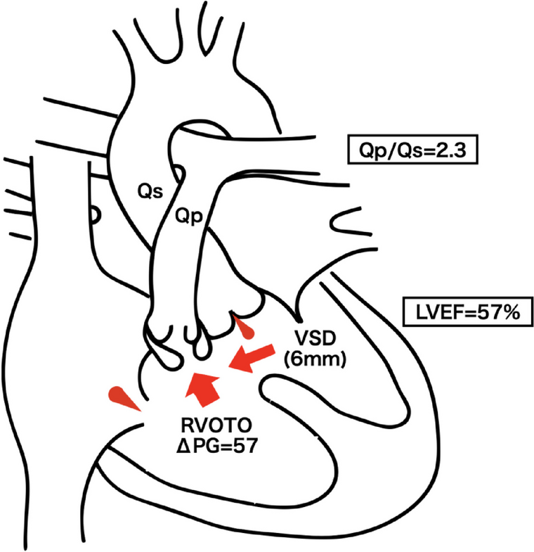
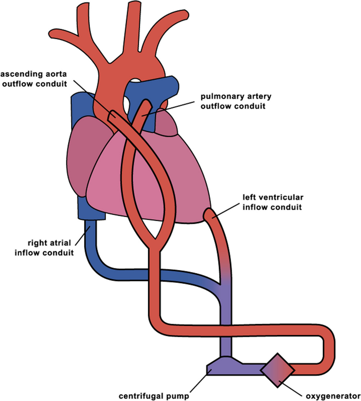
Fig. 1
Central extracorporeal life support system with left ventricular apex vent and pulmonary artery return. A central extracorporeal life support (ECLS) system can be established for patients with severe heart failure due to respiratory failure. The process begins with the introduction of a drainage cannula into the left ventricular apex and connection of the return cannula, which goes to the ascending aorta, to a centrifugal pump circuit equipped with an artificial lung. Given that drainage from the left ventricular apex is often insufficient to maintain total flow in such patients, a peripheral venous cannula is inserted via the femoral vein into the superior vena cava–right atrium (SVC-RA) junction. Subsequently, the cannula is connected to a left ventricular drainage cannula using a Y-connector. To prevent the formation of a left ventricular thrombus, another return cannula is placed into the main pulmonary artery when the drainage volume from the left ventricular apex is exceedingly low. The cannula is connected to the ascending aortic return cannula using a Y-connector. The balance of flow between the two cannulas is controlled by using an adjustable clamp and a separate flow sensor located on one of the outflow tubes
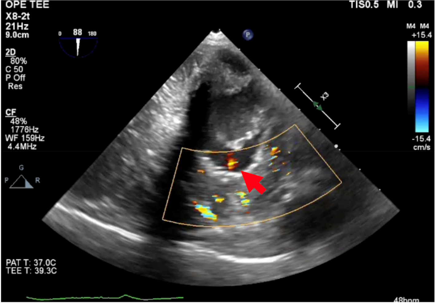
On the second postoperative day in the ICU, the pump flow intermittently decreased, triggering frequent low-flow alerts. At the time, the pump parameters were set at 5440 rpm, and the systemic flow was 2.3 L/min/m2. The external cannula showed no abnormalities such as torsion. The patient remained deeply sedated, showing no signs of asynchrony with the ventilator. An ECG revealed normal sinus rhythm with a heart rate of 105 bpm. The mean arterial pressure, pulse pressure, mean pulmonary arterial pressure, and central venous pressure (CVP) were 65, 5, 19, and 18 mmHg, respectively. Based on these observations, we suspected an abnormal placement of the inflow/outflow cannula. Although transthoracic echocardiography was performed, it failed to produce sufficient images to facilitate a diagnosis, and therefore, the cause could not be determined. Prompt bedside transesophageal echocardiography (TEE) revealed that the anterolateral papillary muscle was suctioned to the vent cannula at the LV apex during systole (Additional file 1: Video 1-pre). This suction was presumed to be a consequence of a reduction in the size of the left ventricular cavity due to diuresis to improve pulmonary congestion and oxygenation. The patient was transferred to the operating room for repositioning of the LV apex cannula under cardiac arrest. Considering that the PVR was less than 3.0 Wood units at the time of weaning from cardiopulmonary bypass, conversion to a BiVAD with an oxygenator was performed according to the standard protocol of our institution. The centrifugal pumps were set to approximately 3500 rpm on both sides, resulting in a pump flow of 4.9 L/min (2.8 L/min/m2). TEE showed the absence of left ventricular apical suctioning (Additional file 1: Video 1-post). Postoperatively, there were no low-flow alarms, and the pump flow remained stable. The patient was weaned off BiVAD 12 days after ICU admission and transferred to another facility 34 days after ICU admission. The patient was finally discharged in a stable condition and without any neurological deficits. She continues to be monitored as an outpatient.
留言 (0)