We evaluated the AH as a predictive factor for impacted stones. The AH was identified as the best predictive factor among hydronephrosis-related factors when compared by AUC analysis. Moreover, the UWT and the AH were preoperative factors to predict impacted stones in multivariate analyses. As risk factors (AH, UWT) increased, there was increase in the rate of patients with impacted stones.
Some predictive factors for impacted stones have been reported, among which UWT is a frequently reported factor [1, 2, 7, 11, 12]. Sarica et al. showed that higher UWT decreases the success rate of shock wave lithotripsy, because UWT is closely related to the degree of impaction [11]. High UWT was shown to be associated with the presence of ureteral edema, polyps, white lesions, stone fixation, longer operation time, and lower endoscopic stone-free rate [7]. The UWT was also determined as a predictive factor for the success of ureteral stent insertion with obstructing ureteral calculi [20]. Impacted stones cause inflammation in the ureteral mucosa and fibrosis of the interstitium. This chronic inflammation can lead to thickening of the ureteral wall, mucosal edema, polyps and stone fixation. Poor endoscopic images make it difficult to disintegrate and remove stones performing URSL. Intraoperative complications were, therefore, reported to be high in impacted stones [6]. In this study, the risk of ureteral injury was higher in patients with impacted stones compared with in patients with non-impacted stones (7.4% vs 0.9%, P = 0.03). The UWT was also determined as an independent predictive factor of impacted stones.
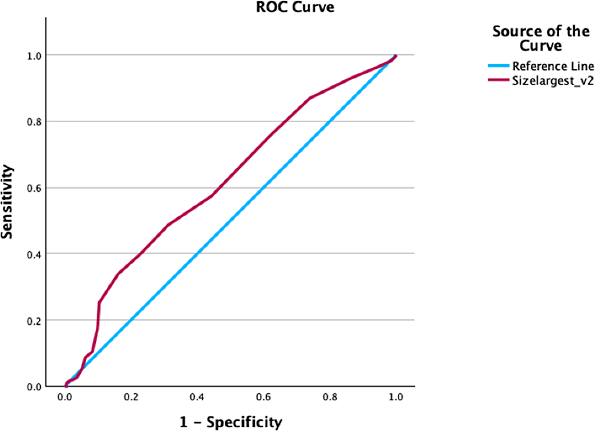
In the case of impacted stones, the degree of hydronephrosis is expected to worsen as the degree of obstruction increases because the impacted stone can strongly obstruct the ureter. Hydronephrosis had been reported as a predictive factor of impacted stones [2, 12, 13]. However, these previous papers assessed hydronephrosis qualitatively using a grading system, and there have been no reports in which hydronephrosis was assessed quantitatively. A problem with evaluating hydronephrosis by grading system is inter-observer error. Han et al. reported lower Kappa values for SFU grades 2 and 3 compared with Grade 1 and 4 when comparing hydronephrosis by renal ultrasound images [21]. The difference between SFU grade 2 and 3 is whether all renal calyces are dilated, but judgement of dilation is dependent on the observer, leading to lower agreement rate [16]. The AH, meanwhile, can be evaluated quantitatively. In the present study, we believe that the predictive ability was high because the dilation of renal calyces can also be quantitatively evaluated. An important issue with measuring the AH is that NCCT coronal images alone cannot evaluate multiple renal calyces because hydronephrosis is evaluated in a one-slice image (Fig. 1). However, by correcting for the long axis of the kidney as in the present study, multiple renal calyces can be evaluated in a one-slice image, and the AH can be measured more accurately. This method, therefore, enabled us to predict for impacted stones. To the best of our knowledge, this is the first report to quantitatively evaluate hydronephrosis in NCCT when predicting impacted stones. RPW, one of the hydronephrosis-related factors, was significantly different between patients with and without impacted stones when comparing patient characteristics, but was inferior to the AH in AUC analyses. RPW could evaluate dilation of the ureter and reflect the degree of impaction, but the RPW could not be used to evaluate dilation of renal calyces, and this would be the cause of inferiority to the AH.
The AH and the UWT were the independent predictive factors, and the number of predictive factors were associated with the risk of impacted stones (Fig. 3). A positive predictive value of 58.2% is certainly not high, but the negative predictive value was as high as 100%. This risk classification is also useful in terms of ruling out impacted stones. If it can determine that a patient does not have impacted stones, it is more likely that the surgery can be performed safely. Moreover, if impacted stones can be predicted, it may be possible to plan certain treatment strategies preoperatively. Anan et al. showed that preoperative percutaneous nephrostomy would improve the stone-free rate in patients who underwent URSL for impacted stones [14]. Other papers also revealed that antegrade URSL in large impacted upper ureteral calculi was a safe and efficient treatment option [9, 10]. It could also explain that the patient may require multiple treatments if there is a high risk of impacted stones. For these reasons, the ability to preoperatively predict impacted stones is very important for clinicians.
This study has some limitations. First, it was retrospective design and was based on a relatively small cohort in a single institution. In the future, the number of cases should be increased and external validation will be carried out to confirm whether the present study is correct. Second, the AH was evaluated by one slice image. Different slices are expected to measure differently. Ideally, if the volume of hydronephrosis could be measured, it would be possible to assess the whole hydronephrosis precisely, but this would likely take a lot of time. In this study, an AH measured by modified coronal image of NCCT took just a few minutes, and it would be useful in daily clinical practice. If 3D software becomes available in the future that can easily measure the volume of hydronephrosis, it would be useful to compare 2D and 3D hydronephrosis. Third, it is expected that there is some inter-measurer error. Demonstration of the reproducibility of this measurement method is therefore necessary, but the AH and RPW are simple and easy to measure and reproducibility is expected to be high. Fourth, the definition of impacted stones was based on the endoscopic findings. Several definitions of impacted stones have been reported, but there is not yet a fixed definition. Although the definition of a guidewire passage in the first attempt is often used, it is difficult to evaluate retrospectively. Therefore, similarly to other reports, we defined impacted stones based on the endoscopic findings [2, 6]. Despite these limitations, the AH and the UWT were considered to be useful predictive factors for impacted stones.


留言 (0)