NIR laser effect on cancer cell lines activity by using Fe3O4@Cu@SiO2 core–shell nanoparticles
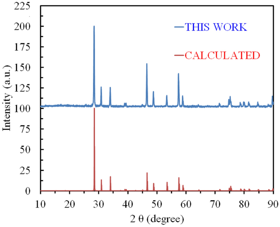
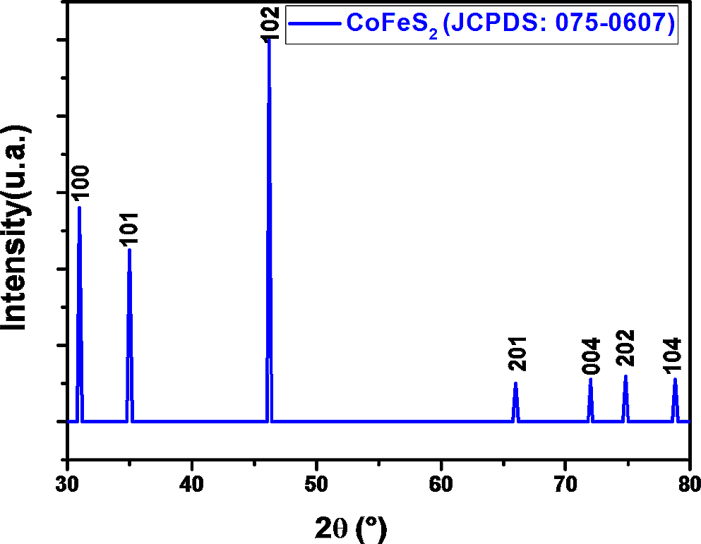
Core–shell structures provide significant advantages in biomedicine, including enhanced stability, controlled release, biocompatibility, multifunctionality and diagnostic capabilities, indicating their potential application in improving drug delivery, diagnosis and therapy. Developing core–multishell nanoparticles with specific sizes, structural properties and optimal absorption characteristics through straightforward methodologies remains a challenge. In this research, we addressed this challenge by employing a three-stage synthesis approach to achieve size control and the ability to fine-tune the properties of Fe3O4@Cu@SiO2 CSNPs. Field-emission scanning electron microscopy was used for microscopic analysis, which verified that Fe3O4@Cu@SiO2 CSNPs had a relatively small particle size of approximately 19.8 nm. In addition, X-ray diffractometer analysis revealed an average crystalline size of 27.54 nm. The nanoparticles demonstrated a stability of approximately − 31.16 mV, indicating their ability to maintain a stable state. Furthermore, the synthesis process involved the incorporation of magnetic and optical properties, which enhanced their biocompatibility. Furthermore, when CP-3 cell lines were exposed to Fe3O4@Cu@SiO2 CSNPs under near-infrared (NIR) laser irradiation, sufficient heat was generated, thereby increasing cell death rate, where the inhibition rate reached 82.44% at the highest concentration and 71.71% at the highest concentration without the use of NIR laser irradiation.

留言 (0)