
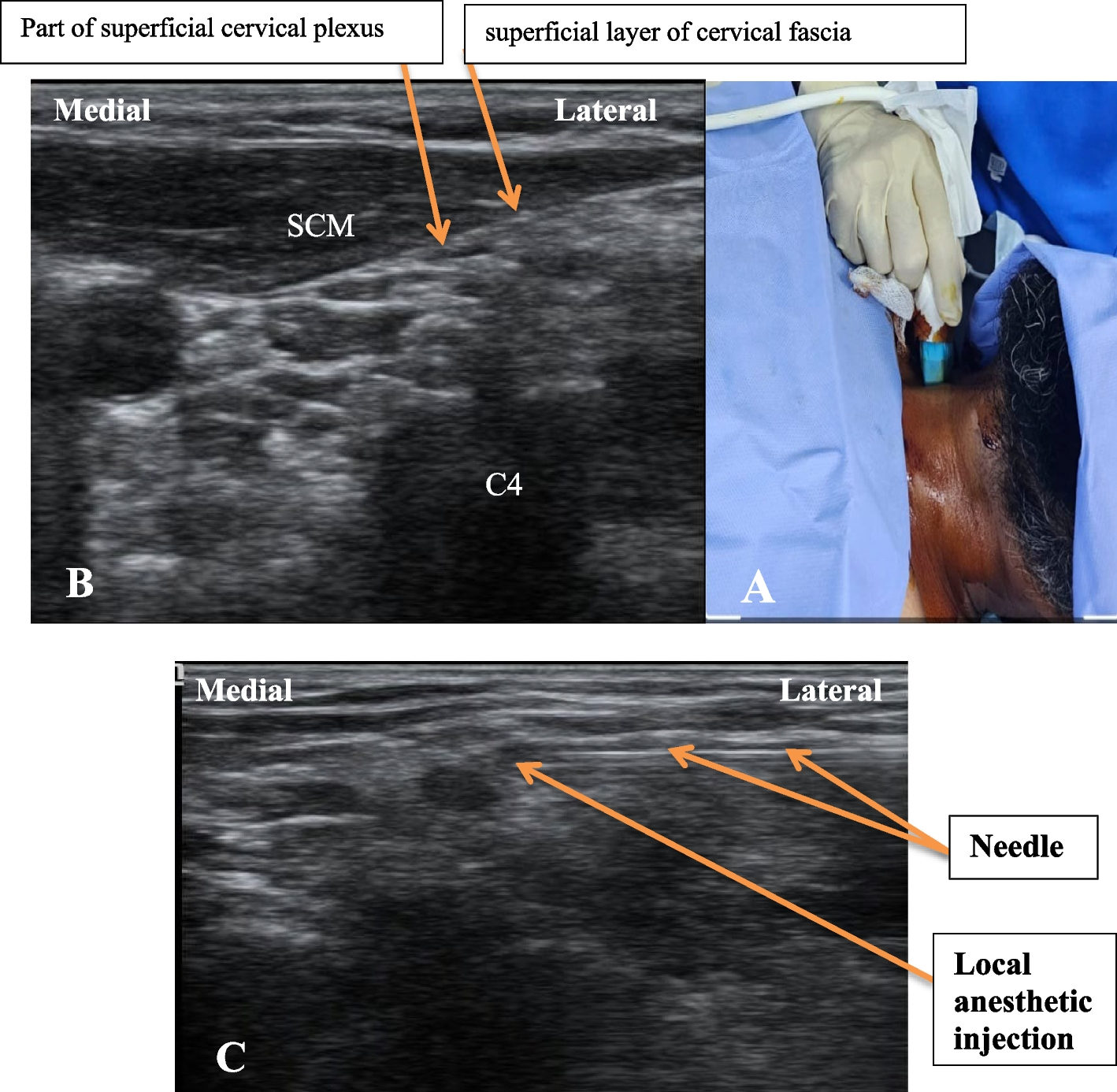
記住我


Between December, 2016 and April, 2018, a total of 100 consecutive patients meeting the inclusion criteria were considered eligible and enrolled in this study. After review of all echocardiographic measurements, 84 patients contained complete, accurate and verifiable data measurements. 13 patients were not included because complete raw data was missing, specifically e’ or E measurements. Two patients were excluded from the final analysis due to poor quality waveforms and one patient was excluded due to a recording error. Given the study design, power analysis could only be performed retrospectively. For a power of 0.95 and α = 0.05, the estimated sample size was n = 31, confirming this analysis was adequately powered for the primary variable.
Demographic and clinical characteristics are summarized in Table 1. The patients in this study could be categorized into four groups: elective valvular replacement or repair, with or without coronary artery disease. Patients had a spectrum of disease pathology, including heart failure, isolated valvular pathology and valvular pathology with coronary artery disease necessitating coronary artery bypass grafting (CABG). None of the patients in this study group had severe mitral stenosis, significant mitral annular calcification or lateral wall motion abnormalities. Data was collected from 27 females and 57 males. Patients had a median age of 66 [60, 73] years and median BMI of 28 [25,32] kg·m-2. The majority (73) were American Society of Anesthesiology (ASA) Physical Status (PS) 4. Ten patients were ASA PS 3 and one was ASA PS 2.
Table 1 Demographic and clinical characteristicsStatic pressures and dynamic hemodynamic variables recorded before and after the fluid bolus are summarized in Table 2. The distribution of these measurements was evaluated for normality. Only the cardiac output (CO) data was normally distributed so the Wilcoxson matched-pairs rank sign test was used for all paired comparisons. All of the measured parameters, except CO, demonstrated statistically significant changes. However, only the parameters associated with the increase in preload demonstrated changes that could be considered clinically significant. The central venous pressures (CVP) increased by 47% from 7.0[5.0, 9.0] to 10 [ 7.0,12.0] mmHg following the fluid bolus. The pulmonary arterial pressures (PAP) increased by 10%, 26% and 17% for systolic (24 [12,31] to 26 [22,34] mmHg), diastolic (11 [9,14] to 14 [11,17] mmHg) and mean (17 [14,19] to 19 [16,23] mmHg) pressures respectively. The stroke volume variation (SVV) decreased by 24% from 8 [7,11] to 6 [4,8].
Table 2 Hemodynamic measurementsPrimary echocardiographic data included lateral mitral annular velocity (e’), peak early ventricular diastolic filling velocity (E) and late filling velocity (A). The distributions of these measured LV functional parameters were evaluated for normality. Only the baseline values of e’ passed the normality test so the Wilcoxson matched-pairs rank sign test was again used for all paired comparisons. The average baseline e’ was 7.8 ± 2.0 cm/s (7.6 [6.6,9.2]) and 8.1 ± 2.4 (7.9 [6.5, 9.6]) following the fluid bolus (p = 0.10). In contrast, E increased from a baseline value of 63 ± 18.4 cm/s (61 [53,73]) to 66 ± 20.5 (63 [53,78]) following the fluid bolus (p = 0.001). Consequently, the E/e’ ratio increase from a baseline value of 8.7 ± 3.6 (7.9 [5.9,11]) to 9.6 ± 4.7 (8.7 [6.4,11]) was also statistically significant (p = 0.0002) (Fig. 2). The average baseline late filling velocity (A) was 59 ± 21.9 cm/s (56 [43.70]) and 61 ± 21.3 (59 [41,79]) following the fluid bolus (p = 0.69) (Table 3).
Table 3 Echocardiographic measurementsFig. 2
(A) Box plot (median, 25%-75% inter-quartile range, whiskers minimum/maximum) and individual patient values of e’ (lateral mitral annular early diastolic tissue velocity) before and after the administration of an intravenous fluid bolus (500 ml). (B) Box plot (median, 25-75% inter-quartile range, whiskers minimum/maximum) and individual patient values of E (early trans-mitral blood flow velocity) before and after the administration of an intravenous fluid bolus (500 ml). (C) Box plot (median, 25-75% inter-quartile range, whiskers minimum/maximum) and individual patient values of E/e’ ratio before and after the administration of an intravenous fluid bolus (500 ml)
All patients were categorized before and after the fluid bolus by their diastolic function grades based on the Swaminathan algorithm [6]. The overall distribution of diastolic function grades did not change following fluid administration (chi square analysis; χ2 = 1.45, df = 3, p = 0.69) (Fig. 3).
Fig. 3
Distribution of diastolic function grades before (pre) and after (post) an intravenous fluid bolus of 500 ml
However, there were many individual patient differences. Of the 35 patients (42%) with changes, 22 patients had worsening functional grading, 20 by one grade, 1 by two grades and 1 by three grades. 11 patients who were normal or grade 1 shifted to grade 2 or grade 3. When compared to patients whose functional assessment did not change, these patients had a greater baseline e’ (8.4[7.4,10] vs. 7.1[6.0,8.1], p = 0.009) but comparable baseline E/e’ (7.7[5.9,9.8 vs. 8.1[6.0,11], p > 0.99). All demographic and baseline hemodynamic variables were also comparable between these two groups. The change in e’ was greater in this group of patients (-0.7[-2.2,-0.03] vs. 0.3[-0.2,0.9], p = 0.003) and the change in E/e’ was statistically greater (2.1[1.0,3.7] vs. 0.6[-0.5,1.6], p = 0.003). The changes in all hemodynamic variables were comparable between these two groups. 13 patients had improved functional grading, 10 by one grade and 3 by two grades. 5 patients who were grade 2 or grade 3 shifted to normal or grade 1. When compared to patients whose functional assessment did not change, these patients had a comparable baseline e’ (7.9[6.5,9.5] vs. 7.1[6.0,8.1]), p = 0.44) and comparable baseline E/e’ (7.5[5.2,10] vs. 8.1[6.0,11], p > 0.99). Demographic and baseline hemodynamic variables were also comparable between these two groups, with the exceptions of age (56 ± 14 vs. 67 ± 11, p = 0.04), diastolic arterial pressure (67 ± 7.4 vs. 59 ± 13, p = 0.02 and heart rate (79 ± 15 vs. 66 ± 12, p = 0.01). The change in e’ was greater in this group of patients (1.7[0.9,2.7] vs. 0.3[-0.2,0.9], p = 0.02) and the corresponding decrease in E/e’ was also greater (-0.8[-2.7,0.4] vs. 0.6[-0.5,1.6], p = 0.03). The changes in all hemodynamic variables were comparable between these two groups, with the exception of heart rate (-12 ± 12 vs. -2.3 ± 9.7, p = 0.04). These individual patient grade changes are summarized in Fig. 4.
Fig. 4
Summary of individual patient changes in diastolic function assessment. Baseline assessments are represented by the total column height. Assessments after a volume infusion are summarized by the shading within each column
The E/e’ values were analyzed to further evaluate the grade assessment shifts between normal/grade 1 and grade 2/grade 3, There was no correlation between baseline vales of E/e’ and the change after fluid administration (Pearson r = 0.02 [-0.19, 0.24], p = 0.8). However, 11 of the 53 patients with a baseline value of E/e’<9 increased to > 9 after fluid administration and the majority (8) of these patients had baseline vales between 7.5 and 9. Only 2 of the 31 patients with baseline values of > 9 decrease to < 9.
留言 (0)