
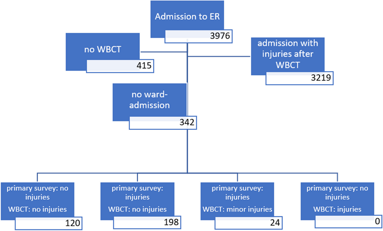
記住我


Of the 31 patients who were followed up and treated in our clinic for traumatic pancreatic and/or duodenal injuries, 18 patients from Erzurum were admitted to our clinic, and 13 were admitted from neighboring cities. Seven were female, and 24 were male. The patients’ mean age was 9 (0–17) years. The patient age group distribution is presented in Table 1.
Table 1 Distribution of demographic, clinical, laboratory, and prognostic data of our pediatric pancreatic/duodenal trauma patient seriesTrauma types experienced by our pediatric patientsThe most common cause of injury was falling from a bicycle, with 12 cases, followed by traffic accidents and collisions, with 7 cases each (Table 1). Handlebar marks were detected on physical examination in 5 of the 12 patients who presented to the hospital after bicycle accidents.
Accompanying injury sitesConcomitant organ injuries accompanied pancreatic/duodenal trauma in eighteen cases. While liver injury was the most prevalent comorbidity of duodenal trauma, pancreatic trauma was most commonly accompanied by lung injury. The distribution of concomitant organ/system injuries is shown in Table 1.
Duodenal and pancreatic injury sitesAnatomically, the most common duodenal injury site was the second part of the duodenum (n = 4), followed by the first part (n = 3) and the third part (n = 1). The pancreatic body (n = 9) and the pancreatic tail (n = 9) were the most prevalent pancreatic injury sites, followed by the pancreatic head (n = 5). In four cases, both pancreas and duodenum injuries were simultaneously observed. Among these, two cases were included in the duodenal injury group because the primary injury was to the duodenum, whereas the other two cases were assigned to the pancreatic injury group since the primary injury was to the pancreas.
Diagnostic stageLaboratory resultsSerum amylase, AST, ALT, and hemoglobin levels were measured at admission in all pancreatic and/or duodenal trauma patients. The mean serum amylase, AST, and ALT values in the case group were significantly high (Table 1). On the other hand, serum amylase values at the time of hospital admission were within the normal range in 3 patients with pancreatic trauma and 2 with duodenal trauma. When the mean serum amylase values obtained at initial admission to the hospital were analyzed, it was noted that the mean serum amylase value of the Grade I pancreatic trauma case group was lower than those of the higher-grade patient groups (Table 2). However, a statistical conclusion could not be achieved because the number of cases was small. A similar suggestion is valid for duodenal trauma even though differences among the grades were present.
Table 2 Number of pancreatic/duodenal trauma cases by grade, mean discharge times, mean serum amylase levels at initial presentation, and mean duration of amylase level elevationImaging resultsAn upright postero-anterior abdominal radiogram was obtained in all patients. Twenty patients underwent FAST, and 30 patients underwent abdominal tomography. When the admission abdominal radiographs of the patients treated for duodenal injury were analyzed, perforation was found in only one patient. In addition, extravasation was found in one patient, free air under the diaphragm in two patients, and duodenal hematoma in one patient in passage radiographs obtained during follow-up.
FAST was performed in 15 patients with pancreatic trauma at admission to the hospital. All 23 patients with pancreatic trauma underwent abdominal CT at admission, whereas FAST was performed in 5, and abdominal CT was performed in 7 duodenal trauma patients. Five of 8 duodenal trauma patients and 3 of 23 pancreatic trauma patients underwent passage radiography due to suspicion of intestinal perforation and absence of clinical improvement. Abdominal MRI was used in five patients with pancreatic trauma during their treatment, and a pancreatic pseudocyst was detected in three of them. Various direct X-ray, CT, and MRI images of the pancreatic/duodenal trauma cases are presented in Figs. 1 and 2.
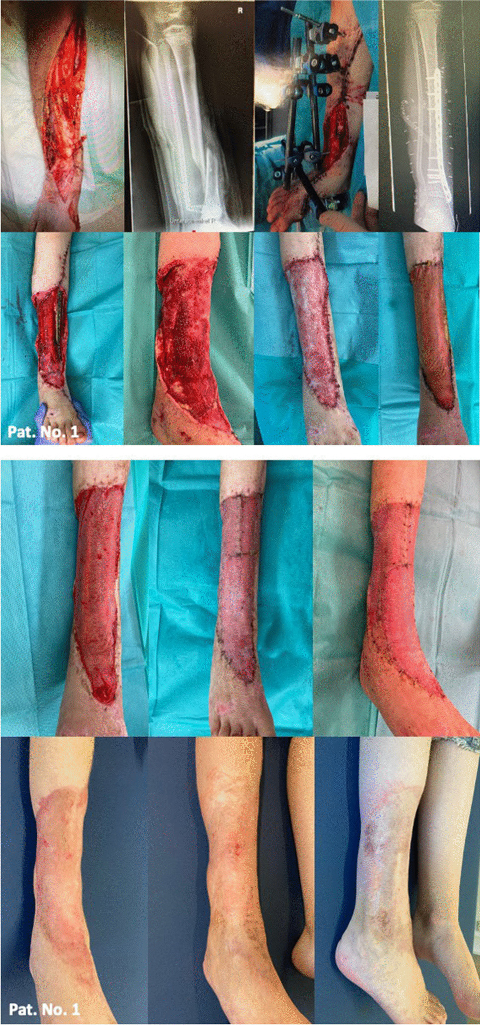
Fig. 1
Various CT and MRI images of the pancreatic/duodenal trauma cases. a Pancreatic laceration on abdominal CT (arrow); b pancreatic laceration on abdominal MRI (arrow); c pseudocyst image on abdominal CT (arrows); d image of free air belonging to duodenal perforation on abdominal CT (arrow); e duodenal hematoma on abdominal CT (circle); f impaired duodenal wall integrity and free air on abdominal CT (arrows)
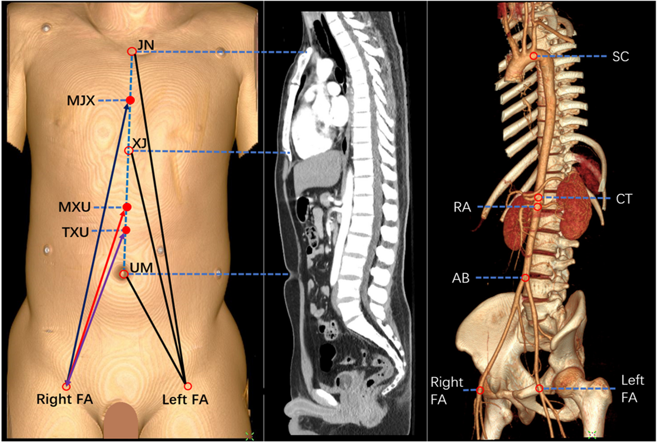
Fig. 2
Various radiological images of pancreaticoduodenal trauma cases. a Duodenal perforation, free air under the right diaphragm on a direct radiograph (arrow). b The pseudocyst’s image on abdominal MRI (arrow). c Duodenal hematoma filling the lumen on gastric-duodenal contrast radiography (arrow). d Direct anteroposterior and lateral X-ray images of the patient with pancreatic trauma due to gunshot injury (radiopaque pellets are observed)
Abdominal CT was superior to FAST in diagnosing or raising suspicion of pancreaticoduodenal traumas when the assessments performed at admission were analyzed (Figs. 3 and 4). An endoscopy was performed on one patient due to accompanying upper GI bleeding; however, the endoscopy revealed normal findings.
Fig. 3
Radiologic features of abdominal CT and FAST in pancreatic trauma patients
Fig. 4
Radiologic features of abdominal CT and FAST in duodenal trauma patients
In pancreatic trauma, diagnosis was made at the time of admission in 21 of 23 cases, whereas within 24 h of admission in one patient, the duration was unidentified in one patient. The diagnosis of duodenal trauma was made within 24 h in four patients, within 48 h in three patients, and within 1 week in one patient (delayed presentation). Overall, the diagnostic process of duodenal trauma was delayed by an average of 2.1 days from the moment of trauma.
Therapeutic managementPancreatic traumaOf the 23 patients who were followed up for pancreatic trauma, 7 were operated on. Of the 13 Grade I pancreatic traumas, one case underwent percutaneous drainage because of a subsequently developing pseudocyst, and another case was diagnosed during laparotomy for gunshot injury and followed up with drain placement. One Grade II case was treated conservatively. Four of the nine Grade III cases were successfully managed conservatively, whereas five patients underwent percutaneous cyst drainage. Two of these patients benefited from the procedure, whereas a Roux-Y cystojejunostomy procedure was performed in one patient who did not benefit from percutaneous cyst drainage. The open surgical procedure achieved cyst drainage and necrotic pancreatic tissue debridement in the remaining two cases. Drains were placed in all patients who underwent surgical procedures.
Duodenal trauma – surgeryAmong 8 duodenal trauma patients, seven were diagnosed with duodenal perforation and operated on. Four patients underwent primary repair + omentopexy only, whereas one underwent primary repair + omentopexy and gastrostomy, one underwent duodenoduodenostomy, and one underwent duodenoduodenostomy with gastrojejunostomy. Drains were placed in all patients who underwent surgical procedures.
Duodenal trauma – conservativeOne patient was diagnosed with duodenal hematoma and was treated conservatively; no operation was planned since the size of the duodenal hematoma did not increase, flow on gastric decompression was reduced, and the patient tolerated nutrition. In the other two Grade 1 duodenal injuries accompanying pancreatic injury, duodenal wall abnormalities were detected and treated conservatively.
Clinical course and outcomeSerum amylase levels were monitored during inpatient treatment and after the patients were discharged. There was no bile leakage in the follow-up of the anastomosis and primary repairs with a drain inserted, but two patients developed brid ileus after discharge. All patients were given prophylactic antibiotics starting from admission. While hospitalized with duodenal trauma, one patient developed candida sepsis, and another developed a fever secondary to pleural effusion.
Long-term follow-up of patients with pancreatic/duodenal trauma was performed with nasogastric drainage. Especially in duodenal trauma, patients were not fed for a long time, and anastomosis lines were not strained with the help of TPN; thus, TPN was started in ten patients to help avoid potential surgical complications. Four patients required blood transfusions (Table 1).
Pseudocyst developmentPseudocysts developed in 11 patients with pancreatic trauma, six of them necessitating surgical interventions. The timing of pseudocyst surgery was 2, 11, 22, 23, and 60 days post-admission in five of them, respectively. The sixth patient’s surgery timing data was unreachable. One patient who had undergone percutaneous cyst drainage experienced re-development of pseudocyst.
Octreotide use in pseudocyst developmentRegarding octreotide use in pseudocyst, four of the patients with pancreatic trauma were started on octreotide, a somatostatin analog, and the common features of the patients were the presence of a concomitant pseudocyst, prolonged elevated amylase levels for more than 30 days, and prolonged hospitalization. Two of these three Grade III patients underwent surgery, and the amount of postoperative drainage decreased with octreotide use. In the other patient with Grade III pancreatic trauma who was started on octreotide, the pseudocyst diameter decreased from 8 to 3 cm. Likewise, the pseudocyst diameter of the Grade I pancreatic trauma patient started on octreotide decreased.
The mean discharge time was 19.57 (2–123) days in pancreatic trauma patients, whereas 18 (7–35) days in duodenal trauma patients. All patients were discharged after the termination of their treatments (Table 1).
留言 (0)