
記住我


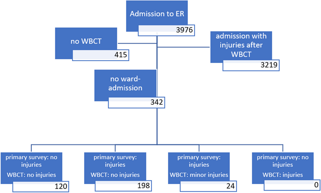
Overall, 213 patients with TBI and intracranial pathology in cranial CT were admitted to our neurosurgical department between 1st of January 2020 and 30th of June 2022. Their distribution across clinical pathways is shown in Fig. 1. Of these 213, 78 patients (36.6%) underwent immediate surgery. Another 12 patients (5.6%) did not receive any further treatment other than the best supportive care as was decided in consensus by the medical team and relatives. Thus, 123 (57.7%) patients were monitored clinically in an intensive care setting. The baseline characteristics of patients that underwent immediate surgery and patients that were monitored are displayed in Table 1.
Fig. 1
Treatment pathways. A total of 213 patients presented to the emergency room and were admitted to neurosurgery. After the first cranial CT, 78 were treated surgically, while 123 were monitored and had a CT6h. Therapy was limited in 12 patients (“Drop-Out*”). Nine of 123 monitored patients showed clinical deterioration, had emergency imaging before CT6h, and went on to surgery. The remaining 114 patients had CT6h, eight of which went on to have surgery that was planned after first CT already in seven cases (see text). A total of 106 patients received further conservative treatment after CT6h, nine of which showed later deterioration or failure of conservative treatment and went on to surgery after days to weeks. Ninety-seven of 213 patients received definitive conservative treatment
Table 1 General characteristics of patient cohort. Left column: parameters, age in years, sex (male:female), Glasgow coma scale, neurological deficits. Middle column: patients that had surgery after the first CT (n = 78). Right column: the remaining patients that initially received conservative treatmentIn summary, mean age (67.6 ± 18.7 for immediate surgery and 64.0 ± 20.2 for initial conservative treatment) and sex distribution (male:female 1.8:1 for immediate surgery and 1.7:1 for initial conservative treatment) were similar. While the immediate surgery group showed a higher ratio of patients with GCS 3–8 (18/78 or 23.1% vs. 6/123 or 4.9%), most patients were GCS 13–15 in both groups (56/78 or 71.8% in immediate surgery vs. 105/123 or 85.4% in initial conservative treatment). Eight of 135 (5.9%) cases in the group Surveillance + Drop Out had a GCS of 3 to 8. These patients did not undergo invasive ICP monitoring for the following reasons: Of these eight, four were dropouts with no further treatment intended. Three were postictal and showed rapid clinical improvement. In one case, the patient showed a small SDH of only 3 mm, while alcohol blood levels indicated severe intoxication.
However, the immediate surgery group displayed neurological deficits more than twice as often (45/78 or 57.7% vs. 28/123 or 22.8%), motor deficits six times more often, and aphasia five times more often than initially conservative patients, whereas behavioral changes (12.2%) were the most prevalent changes in patients initially treated conservatively. In both groups, “fall due to tripping” was the most common trauma mechanism (67.9% in immediate surgery, 65.9% in initial surveillance), while fall from stairs (1.3% in surgery vs. 9.8% initial conservative treatment) and traffic accidents (9.0% in surgery vs. 13.0% initial conservative treatment) predominated in the conservative group. Also, the incidence of coagulation disorders was similar in both groups (53.8% in initial surgery vs. 50.4% in initial conservative treatment).
In patients that were immediately chosen for surgery, 11/78 (14.1%) had epidural hematoma, 65/78 (83.3%) suffered from subdural hematoma, 6/78 (7.7%) sustained traumatic brain contusions, and 14/78 (17.9%) had traumatic subarachnoid hemorrhage (multiple injuries per patient possible). In patients that were initially treated conservatively, 8/123 (6.5%) had epidural hematoma, 80/123 (65.0%) suffered from subdural hematoma, 52/123 (42.3) sustained traumatic brain contusions, and 68/123 (55.3%) had traumatic subarachnoid hemorrhage. Thus, brain contusions were predominantly nonsurgical lesions, while epidural hematomas were evacuated immediately in two-thirds of cases.
One hundred fourteen of 123 patients in the conservative group received CT6h at a median 6 h after first CT, while nine patients underwent earlier CT (mean time to CT 216 min, see Fig. 2) due to neurological deterioration. All nine patients with neurological deterioration underwent immediate surgery, while another eight patients (8/114, 7%) that received the CT6h underwent surgery as well. Of the remaining 106 patients that were transferred to neurosurgical wards or continued to be monitored on the intensive care unit depending on their clinical status, another nine patients (9/104, 8.7%) underwent delayed surgery after two or more follow-up CTs between 25 h and 1 month after admission leaving 97 out of 123 (78.9%) patients initially treated conservatively to definitive conservative treatment.
Fig. 2
Timing of clinical deterioration under intensive care surveillance before routinely scheduled follow-up CT. Nine patients deteriorated ahead of scheduled CT6h and had emergent CT a median of 216 min after first CT. Patients A to G and I showed sole neurological worsening, while patient H needed intubation due to COVID pneumonia and received an external ventricular drain
Impact of CT6h on surgical decision makingOf 123 patients that were initially treated conservatively and monitored clinically, 17 patients were operated after the first follow-up CT (9 had earlier CT due to clinical deterioration, 8 after CT6h). Of note, all patients that deteriorated neurologically (9/17) did so before completion of the 6-h interval and received an earlier CT than CT6h. The timing of clinical deterioration after first CT is displayed in Fig. 2 and shows a clustering between 3 and 4 h after initial CT (mean time to control CT due to clinical deterioration 216 min), while exemplary CT images are displayed in Fig. 3. Eight of nine patients deteriorated in terms of GCS or a new neurological deficit, whereas one patient (patient H in Fig. 3) had to be intubated due to respiratory insufficiency and COVID pneumonia and received an external ventricular drain for intracranial pressure monitoring.
Fig. 3
Examples of first and second cranial CT in patients that deteriorated before CT6h and received emergent imaging. The letters at the top left corner of images correlate with the patient identifier in Fig. 2. All patients that showed early deterioration before CT6h received surgical treatment. Left column: CT at admission, right column: emergent follow up CT. CT, cranial computerized tomography. (For illustrative purpose, CT slices with greatest hemorrhage extension are shown. Therefore, slices of initial and control CT might not be at the same level)
Detailed chart review for the eight patients that were operated on after CT6h revealed that in seven out of eight cases, surgery was indicated after the first CT but scheduled for the following morning usually due to subacute or acute-on-chronic traumatic subdural hematoma. In these cases, CT6h was ordered simply for updating CT imaging before surgery, detected no change, and did not impact the intended treatment. Only in one case (1/114) routine follow-up CT led to a change in treatment protocol since it displayed a vastly different intracranial pathology than initial CT due to poor quality of the initial examination and correction of radiological artifacts.
The nine patients that were operated days to weeks after first and second CT did so due to various reasons ranging from self-dismissal against medical advice (n = 1), delayed cerebral edema after 3 and a half days (n = 1), delayed onset of headache or neurological deficits after several days (n = 4), an initially stable situation under anticoagulation that permitted a conservative treatment attempt with correction of coagulation disorders (n = 1), and a failed conservative treatment attempt in elderly with severe comorbidities and subdural chronification (n = 2). Of note, out of nine patients that received late surgery, eight had a stable CT6h (four times acute subdural hematoma in combination with contusions or traumatic subarachnoid hemorrhage or frontobasal fracture, five times subacute subdural hematoma).
In summary, only one out of 114 CT6h led to a change in surgical decision making due to poor quality of the initial examination. On the other hand, CT6h did not catch a single imminent neurological deterioration. On the contrary, all patients with neurological deterioration under clinical monitoring worsened before completion of the 6-h interval and were detected clinically. The timing of deterioration clustered between 3 and 4 h after initial CT. Additionally, eight out of nine late deteriorations/indications for surgery had an initially stable CT6h. The predictive value of CT6h for change in surgical management was < 1%. The negative predictive value for later surgical therapy after CT6h was 91.5%, meaning that 8.5% of patients without initial surgical therapy suffered from late clinical deterioration or failure of conservative treatment.
Impact of CT6h on surgical decision making in mild traumatic brain injuries without major neurological deficitSince follow-up CT is most controversial in patients presenting with mild TBI (GCS 13 to 15) without any major neurological deficit, they were analyzed separately in this study. Notably, in our clinical routine in an emergency setting, not all patients were screened for subtle neuropsychological deficits. Therefore, only the absence of major neurological deficits can be described by us. One hundred three out of 213 (48.4%) patients presented with an intracranial pathology in initial cranial CT at admission, but with GCS 13 to 15 and without any major neurological deficit. Fifteen out of 103 (14.6%) received immediate surgery due to extension of pathologies alone (including multiple injuries per patient four epidural hematomas, twelve subdural hematomas), and a further six patients (6/103, 5.8%) were operated after CT6h (one EDH, two SDH, one contusion with tSAH, one contusion only, and one tSAH with concomitant depressed calvarial fracture). Again, in half of these patients (3/6), the follow-up CT was carried out earlier than planned due to early clinical deterioration, while in the other three cases, surgery was indicated after the first CT but scheduled for the following morning. In these cases, CT6h was ordered simply for updating CT imaging before surgery, detected no change, and did not impact treatment. However, of the nine patients operated on after days to weeks as described above (see Fig. 1), eight patients presented initially with GCS 13 to 15 without any neurological deficit. Seventy-three out of 103 patients (70.9%) went on to definitive conservative treatment (the remaining 9 patients dropped out due to limitation of therapy by family or patient’s will). This confirms that no imminent deterioration was anticipated by CT6h. Further, it demonstrates that even patients with mild TBI without any major neurological deficit but intracranial pathology have a risk for surgically relevant pathology (21/103, 20.4% after first follow-up CT) and clinical deterioration necessitating monitoring.
Impact of coagulation disorders on intracranial injury at admission and follow-upAs shown in Table 1, management of coagulation disorders is increasingly relevant in neurosurgery since approximately 50% of patients with TBI and intracranial pathology present with coagulation disorders. Approximately 38% of patients presented to the emergency department with a prior medical history of anticoagulation medication. In our cohort, acetylsalicylic acid (40/213, 18.8%) and direct oral anticoagulation drugs were the most common anticoagulation medication (27/213, 12.7%) to be dealt with, whereas other antiplatelet drugs (9/213, 4.2%) and vitamin K antagonists (8/213, 3.8%) were less often. Additionally, 32% of patients presented with abnormal laboratory testing to various reasons regularly overlapping with the intake of anticoagulation medication.
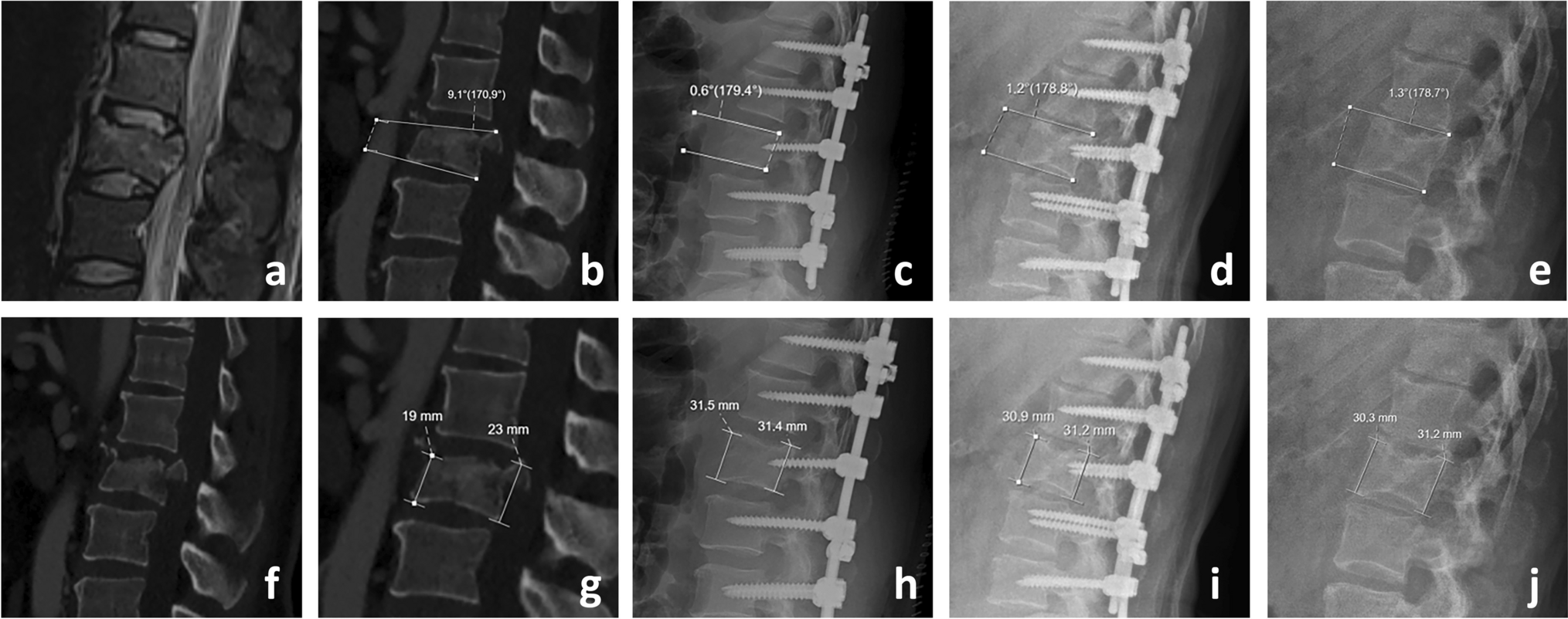
The distribution of coagulation disorders across patients with different injury patterns is displayed in Table 2 and Figs. 4 and 5 (anticoagulant medication, anticoagulant medication, and altered coagulation laboratory testing, altered coagulation laboratory testing without anticoagulant medication, and no detected coagulopathy). Of note, none out of 9 patients that presented with isolated epidural hematoma suffered from a coagulation disorder, highlighting this lesion association with a specific injury mechanism, while eight out of nine patients with isolated traumatic brain contusions (without traumatic subarachnoid hemorrhage) showed a impaired coagulation (4/9 taking anticoagulation drugs, 7/9 displaying abnormal laboratory testing). Patients with subdural hematoma (64.5% with coagulation disorder) and traumatic subarachnoid hemorrhage (45% with coagulation disorder) as well as patients with combined brain injuries (43.9% with coagulation disorders) showed ratios of coagulation disorders approximating 50% as was the mean percentage across all injury patterns. Specific analysis of mean volume (contusions) or mean diameter (subdural hematoma) of intracranial pathology revealed that patients with coagulation disorders showed no significant difference in mean lesion size at presentation in isolated subdural hematoma (23 ± 9.9 mm without coagulopathy vs. 20 ± 9.8 mm with coagulopathy on first CT across all therapeutic groups, p = 0.16) or in subdural hematoma combined with other traumatic lesions (15.7 ± 10.9 mm without coagulopathy vs. 17.7 ± 9.9 mm with coagulopathy p = 0.24) or contusions in combined brain injuries (9.9 ± 15.2 ml with coagulopathy vs. 6.2 ± 11.6 ml without coagulopathy, p = 0.32). Of note, there was no difference in mean lesion size in patients with antiplatelet agents versus anticoagulants.
Table 2 Characterization of coagulation disorders across injury patterns (left) and according to initial treatment (right). Isolated injuries: Cont, contusions; EDH, epidural hemorrhage; SDH, subdural hemorrhage; tSAH, traumatic subarachnoid hemorrhage; Mix, any combination thereof. PC, platelet count; Quick, partial thromboplastin time according to Quick; INR, international normalized ratio; aPTT, activated partial thromboplastin time. ASA, acetylsalicylic acid; DOAC, direct oral anticoagulants; VKA, vitamin K antagonistsFig. 4
Distribution and type of coagulation disorder across injury patterns. Percentage of coagulation disorder due to medication (anticoagulants/antiplatelet drugs), labs (abnormal laboratory parameters without responsible medication), and both in patients with isolated contusions (CONT), epidural hemorrhages (EDH), subdural hemorrhages (SDH), traumatic subarachnoid hemorrhage (tSAH), or combinations thereof (MIX). This graph illustrates that about half of all patients presented with coagulation disorders. While isolated contusions had a high association with coagulation disorders, epidural hematomas are known to be more associated with the trauma mechanism
Fig. 5
Cumulative size distribution for maximal diameter of SDH (A) and volume of contusions (B) in any constellation showed no significant differences between patients that presented with (CD) or without coagulation disorders (no CD) in the first CT scan. Size progression in mm for SDH (C) and ml for contusions (D) between first and second CT was classified into four groups (< − 2 mm/ml: decrease/redistribution, − 2 to + 2 mm/ml: stable, + 2 to + 6 mm/ml: small increase, > + 6 mm/ml: high increase). For patients that showed radiological progression of SDH as part of combined brain injuries under management of coagulopathy (E), those with coagulation disorders (CDs) showed more often higher increase of lesion size compared to patients without CDs (diameter increase > 6 mm: 11.1% vs. 2.8%). This effect was not observed for contusions (F). Please note that patients who were operated after 1st CT do not show up in the graphs C, D, E, and F
Development of lesion size in all patients with subdural hematoma or contusions undergoing initial control imaging is displayed graphically in Fig. 5, while the ratio of patients with relevant increase in lesion size is additionally shown in Table 3. In summary, 16.7% (6/36) of patients with subdural hematoma without coagulopathy showed radiological progression of more than 2 mm increase in hematoma diameter, while 17.8% (8/45) of patients with subdural hematoma with coagulopathy (under institutional standard-of-care clinical management of coagulation disorders) presented this same radiological progression. Of note, while hematomas that showed radiological progression between 2 and 6 mm were predominantly in patients without coagulopathy (13.9% of patients without coagulopathy vs. 6.7% of patients with coagulopathy), an increase of more than 6 mm was mostly in patients with coagulopathy (11.1% of patients with coagulopathy vs. 2.8% of patients without coagulopathy) as displayed in Fig. 5. This effect does not pertain to contusions.
Table 3 Development of lesion size. Left: lesion size at first CT in mm for patients with SDH in any combination of injuries or traumatic brain contusions in any combination of injuries as means with standard deviation in mm for SDH and ml for contusions (no CD, no coagulation disorder; CD, coagulation disorder). Right: development of lesions from first to second CT with ratio of lesions decreasing in size > 2 mm, remaining stable, increase in size of 2–6 mm or more than 6 mm (ml for contusions)
留言 (0)