Posterior cerebral artery (PCA)-accessory PCA (hyperplastic anterior choroidal artery) anastomosis detected on magnetic resonance angiography
Purpose
To describe a case of posterior cerebral artery (PCA)-accessory PCA (hyperplastic anterior choroidal artery) anastomosis detected on magnetic resonance angiography.
Methods
A 76-year-old man with a history of cerebral infarction underwent cranial magnetic resonance (MR) imaging and MR angiography of the intracranial region for the evaluation of brain and vascular lesions. The MR machine was a 3-Tesla scanner. MR angiography was performed using a standard three-dimensional time-of-flight technique.
Results
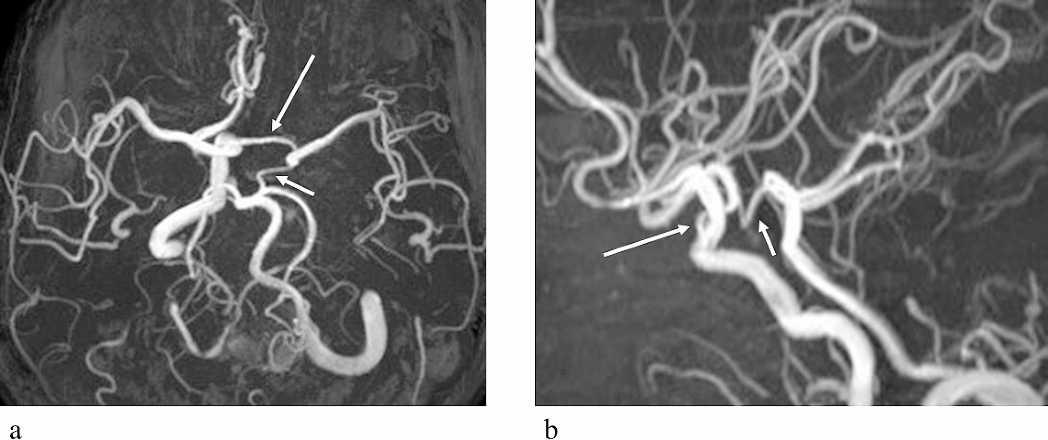
There were two right PCAs. The parieto-occipital and calcarine arteries of the right PCA arose from the right ICA, indicative of accessory PCA, and there were three stenotic lesions at the proximal segment of this artery. The temporal artery of the right PCA originated from the basilar artery. A small anastomotic channel between these two arteries was identified on partial maximum intensity projection (MIP) images. Computed tomography angiography was additionally performed and the findings were confirmed.
Conclusion
We speculated that the pressure gradient between the PCA and the accessory PCA enlarged the anastomotic channel. Partial MIP images are useful for diagnosing small arterial variations using MR angiography.


留言 (0)