Plant materials
M. siamensis flowers were purchased from a Thai traditional drug store in Nakhon Si Thammarat Province, Thailand. The collection of plant materials followed the relevant guidelines and regulations of The Plant Varieties Protection, Department of Agriculture, Ministry of Agriculture and Cooperatives, Thailand. The plant was identified by Assoc. Prof. Tanomjit Supavita, a botanist at the School of Pharmacy at Walailak University. The plant material was deposited at the School of Medicine, Walailak University, with identification voucher number SMD122006002. Plant materials were rinsed with distilled water before being dried in a hot air oven at 60 °C and pulverized to coarse powder using a herb grinder.
General experimental procedure
Silica gel (230–400 mesh, Sili Cycle Inc., Canada) was used for column chromatography (CC). All solvents were purchased from Labscan (Thailand) and were of analytical grade. All reagents were purchased from Sigma-Aldrich (USA). The nuclear magnetic resonance (NMR) spectra were obtained using an Avance NEO spectrometer (Bruker) at 500 MHz for 1H.
Extraction and isolation of compounds
The dried M. siamensis flower powder (8.0 kg) was macerated extensively in dichloromethane (15 L) for 72 h with occasional shaking. The solvent was filtered through Whatman No. 1 filter paper. The residue was re-macerated for 72 h, and this procedure was repeated thrice. The filtrates were combined and concentrated using a rotary evaporator (Rotavapor®, Buchi, China) at 50 °C to obtain a brownish viscous crude extract (76.8 g). Finally, the dried crude extract was kept in screw-cap containers and stored at 4 °C for further use.
Based on its in vitro antiplasmodial activity, HTX was isolated according to our previous study, with some modifications [20]. The dichloromethane extract (75 g) underwent quick CC (QCC) through silica gel by step-gradient elution starting with dichloromethane. The polarity was increased with ethyl acetate (EtOAc) and acetone to yield 12 fractions (F1-F12), based on thin-layer chromatography profiles. Fraction 6 (8.2 g) was purified by QCC using 10% EtOAc in hexane to produce five subfractions (6A-6E). Subfraction 6B (4.13 g) was further purified by QCC using 15% EtOAc in hexane to obtain four subfractions (6B1-6B4). Subfraction 6B2 (2.66 g) was further purified by CC with 15% EtOAc in hexane to obtain compound 1 as a yellow powder (62.6 mg).
Animals
Healthy adult male Institute of Cancer Research (ICR) mice weighing 25–30 g body weight and aged 6–8 weeks were obtained from Nomura Siam International Co., Ltd., Pathumwan, Bangkok, Thailand, for antimalarial activity, acute toxicity, and pharmacokinetic testing. The study was approved by the Animal Ethics Committee of Walailak University (certificate number: WU-ACUC-65015), and in accordance with Ethical Principles and Guidelines for the Use of Animals by the National Research Council of Thailand. All mice were housed in cages under standard environmental conditions at a relative humidity of 50–60% and a room temperature of about 22 °C (± 3 °C) under a natural 12-hour dark-light cycle. Mice had free access to pellet diet and clean drinking water. All mice were acclimatized to the experimental environment for a week before beginning the experiment. This study was reported in accordance with ARRIVE guidelines.
Parasite inoculation
Wild-type P. berghei ANKA strain was obtained from Thomas F. McCutchan (BEI Resources, NIAID, NIH). P. berghei-infected red blood cells were administered intraperitoneally to donor mice. When parasitemia levels reached 20–30%, blood was collected from the heart using a cardiac puncture procedure and placed in a vacutainer heparinized tube for injection into experimental animals [21].
In vivo 4-day suppressive test of P. berghei
A 4-day suppressive test was used to measure the schizonticidal activity of HTX in P. berghei-infected ICR mice. This test was performed according to a previous method, with some modifications [22, 23]. Male ICR mice were randomly divided into six groups, each consisting of five mice, as shown in Table 1. All mice were intraperitoneally injected with 1 × 10⁷P. berghei ANKA-parasitized erythrocytes. Treatment was initiated four hours after the mice were infected with Plasmodium parasite on the first day (D0). The negative control group was administered 150 µl of 2% dimethyl sulfoxide (DMSO) in normal saline solution (NSS), whereas the positive control group was administered 3 mg/kg body weight of artesunate and chloroquine intraperitoneally each day. For each experimental group, animals received daily intraperitoneal doses of 1, 3, or 10 mg/kg body weight of HTX. Mice received each substance daily for 4 days (D0-D3). On the fifth day (D4), parasitemia was determined using Giemsa-stained thin blood smears prepared from the tails of each mouse. Mice were euthanized at the end of the experiment by intraperitoneal injection of pentobarbital (200 mg/kg body weight).
Finally, the percentage suppression of parasite growth was calculated using the following equation:
$$\% }\frac} \hfill \\} \hfill \\ \end }}}}} \times }$$
Table 1 Group classifications and doses used in the 4-day suppressive testAcute toxicity test
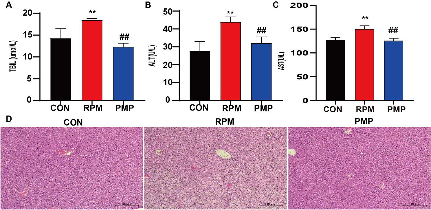
Acute HTX toxicity was determined in non-infected ICR mice aged 6–8 weeks and weighing 25–35 g using the Organization for Economic Cooperation and Development standard criteria [24]. Fifteen mice were randomly divided into three groups (n = 5): untreated, negative control, and HTX-treated. All mice fasted for three hours prior to the experiment, and only drinking water was available. HTX was dissolved in 2% DMSO in NSS at 50 mg/kg body weight. In the treatment group, the first mouse was intraperitoneally given a single dose of 50 mg/kg body weight HTX, whereas in the negative control group, the first mouse was intraperitoneally given 200 µL of 2% DMSO in NSS. After dosing, mice were observed for 24 h for gross physiological and behavioral changes, such as sleep, hair erection, poor appetite, convulsions, diarrhea, lacrimation, salivation, mortality, and other manifestations of toxicity. If death was not observed within 24 h in the first mouse, the same dose was administered to 4 more mice, and they were observed for signs and symptoms of toxicity for 14 days. All mice were anaesthetized using pentobarbital (60 mg/kg body weight) by intraperitoneal injection. Blood samples were collected via cardiac puncture into heparinized tubes for biochemical analyses. The liver and kidney tissues were collected for histological examination. The mice were subsequently euthanized with an intraperitoneal injection of pentobarbital (200 mg/kg body weight).
Histopathological examination
Histopathological examinations of liver and kidney tissues were performed in accordance with standard histological protocols, as reported previously [25, 26]. In brief, all tissues were fixed in 10% buffered formalin, dehydrated using a gradient series of ethanol solutions, cleaned with xylene, and deposited in a paraffin mold. The paraffin blocks were then sectioned to a thickness of 5 μm using a rotary microtome, transferred to glass slides, and stained with hematoxylin and eosin (H&E) solution. The stained slides were examined under light microscopy by two independent observers who were blinded to the experimental groups for examining the histological changes.
In vivo pharmacokinetic studySamples preparation
A pharmacokinetic study was conducted in non-infected ICR mice aged 6–8 weeks and weighing 25–35 g. Male ICR mice were randomly divided into two groups, each consisting of five mice. The first group is a negative control group and was injected with 150 µL of 2% DMSO in NSS, whereas the second group is a test group that was administered 5 mg/kg body weight of HTX compound intraperitoneally. Blood (50 L) was taken from the tail vein prior to administration of test compounds (0 h) and after at, 0.5, 1, 2, 3, 5, 8, 24, 48, and 72 h. Blood samples were collected in microtubes containing ethylenediaminetetraacetic acid (EDTA) and kept on ice until they were centrifuged at 3,000 × g for 10 min at 4 °C to allow the collection of plasma, which was stored at − 80 °C.
Liquid chromatography triple quadrupole mass spectrometry (LC-MS/MS)
LC-MS/MS (Agilent 1290 infinity LC and Agilent 6490 triple quadrupole mass spectrometer) with an electrospray ionization (ESI, Agilent Technologies, USA) source was used to detect HTX in the plasma. Using a VertiSepTM USP C18 column (4.6 mm × 150 mm, particle size = 5 μm; Vertical Chromatography Co., ltd., Nonthaburi, Thailand), the separation was achieved. Acetonitrile:1 mM formic acid (7:3) was used as the mobile phase and was maintained at a constant flow rate of 0.5 mL/min. The injection volume was 1 µL, and column temperature was maintained at 30 °C. The following operating conditions for the MS were optimized: the source temperature was kept at 200 °C, the ion spray voltage for the positive mode was set at 3 KV, and the collision energy for HTX was set at 43 V. Nitrogen was used as the collision gas. The nebulizer and sheath gas flow rates were set to 14 and 11 L/min, respectively. The MassHunter qualitative analysis software was used to acquire the data (Agilent Technologies, Inc. Headquarters, CA, USA). HTX was quantified using the transitions for HTX at m/z 303→199 in the multiple reaction monitoring (MRM) mode.
Preparation of the standard and quality control samples
By dissolving precisely weighed HTX and reference compounds in acetonitrile, stock HTX solutions were prepared. By diluting the stock solutions using a mixture of acetonitrile and water (2:8, v/v), a series of working solutions with concentrations between 7.81 and 1000 ng/mL were prepared. All solutions were stored at 4 °C. By spiking the blank plasma (50 µL) with 25 µL of standard working solutions, HTX calibration standards (0.78, 1.56, 3.13, 6.25, 12.5, 25.0, 50, 125, and 250 ng/mL) were prepared. The extraction solvent ethyl acetate (0.5 mL) was added to 75 µL of HTX-spiked plasma. The sample was vortexed for 1 min, followed by ultrasonic vibration for 15 min. Then, centrifugation for 10 min at 15,000 × g was done. The upper organic layer was transferred to a new microtube and dried via evaporation. The dried extract was reconstituted in 150 µL of 50% (v/v) acetonitrile in water. HTX recovery experiments were performed to confirm extraction efficacy. The effectiveness of liquid-liquid extraction was then determined by comparing the peak regions of the extracted HTX-spiked plasma with those of HTX spiked with blank plasma extract.
Pharmacokinetic analysis of HTX
The plasma concentration-time profiles of HTX were determined by non-compartmental analysis with PKSolver 2.0 software. Pharmacokinetic parameters were calculated to estimate the areas under the concentration-time curve (AUCs) from 0 to 48 h and from 0 to ∞, elimination half-life (t1/2), maximum concentration achieved (Cmax), time to attain Cmax (Tmax), mean residence time (MRT), apparent volume of distribution, and clearance. Additionally, the apparent volume of the central or plasma compartment, apparent volume of the peripheral compartment, transfer rate constant from the central compartment to the peripheral compartment, and transfer rate constant from the peripheral to central compartment were calculated using the two-compartment model.
Statistical analysis
Results were presented as means ± SEM. IBM SPSS Statistics version 23.0 software (SPSS, IL, USA) was used for statistical analysis. Normal distribution was tested using the Kolmogorov–Smirnov goodness-of-fit test. Statistical significance of parasitemia inhibition was determined using one-way ANOVA, followed by Tukey’s multiple comparison test. Statistical significance was set at 0.05 (p < 0.05).


留言 (0)