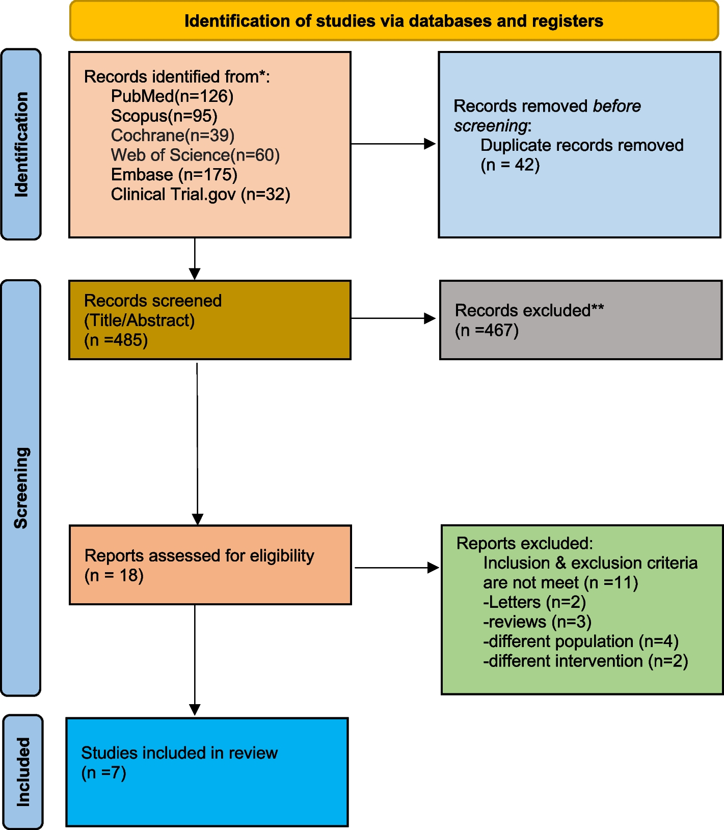
The initial analysis of the PMA randomised clinical trial's findings improves our understanding of the benefits of using PMA with RMA compared to using RMA alone for treating severe ischemic mitral regurgitation. The 5-year results indicate that the combination of papillary muscle surgery and annuloplasty was more effective than standard annuloplasty alone for the treatment of ischemic MR. This resulted in improved ventricular geometry, remodeling, and function. The research uncovered a significant difference between groups in the rank-based evaluation of left ventricular reverse remodeling after 5 years. The findings demonstrated a consistent and substantial enhancement in LVEDD, LV end-systolic diameter, and ejection fraction in the PMA cohort. On the other hand, the RMA alone group displayed progressive ventricular cavity enlargement and LV function decline, indicating inadequate counteraction or prevention of detrimental remodeling that occurred over time in ischemic MR [3].
In this second analysis, our objective is to thoroughly examine how reducing interpapillary distance through surgical approximation of papillary muscles can recover left ventricular geometry whilst mitigating the risk of recurrent MR.
Principal findings
The reappearance of ischemic MR after RMA ± CABG is traditionally thought to occur due to progressive LVEDD enlargement caused by chordal tethering. However, our findings illustrate that progressive LVEDD enlargement does not lead to the recurrence of ischemic MR, as long as the IPMD is surgically stabilized by adding PMA. Possibly, the combination of RMA and PMA provides a solution for both the valvular and sub-valvular mitral apparatus, similarly to MVR with chordal sparing.
PMA affects normal spatial relationships of the components of MV in patients with SIMR
The erroneous assumption that the leaflet tethering in ischemic MR resulted from apical displacement of the papillary muscles was initially made due to the use of tethering lengths (2D parameters) in earlier studies to evaluate the 3D displacement of the muscles [6,7,8,9,10,11,12,13,14]. However, subsequent studies looking at the effect of LVEDD enlargement in ischemic MR with a 3D assessment challenged this notion. Experimental induction of acute ischemic MR did not displace the papillary muscles at the apex. Instead, it restricted the distance between the posterior commissure and the posterior papillary muscle, according to reports [15,16,17]. Moreover, three-dimensional evidence reveals that experimental models studying non-acute ischemic MR yield fitting results. In cases of sub-acute or chronic ischemic MR, an enlarged LVEDD results in decreased mitral annulus to posterior papillary muscle distance compared to the control group (3.16 ± 0.41 cm vs. 3.82 ± 0.53 cm respectively, p = 0.01). This change in shape resulted from two different forces affecting the posterior papillary muscle—a force towards the back (0.58 ± 0.4 cm compared to 1.39 ± 0.46 cm in the control and ischemic groups, p < 0.01) and a force from the side (0.66 ± 0.55 cm compared to 1.19 ± 0.70 cm in the control and ischemic groups, p < 0.01). On the other hand, the ischemic enlargement of the LVEDD did not significantly shift the anterior papillary muscle. Hence, 3D data contradicts the idea that ischemic MR is caused by displacement of the apical papillary muscle and points instead to displacement of the posterior papillary muscle in the posterolateral region as the primary cause. This hypothesis is supported by studies that have attempted to rectify the sub-valvular vectors. Balloon repositioning of a posterior papillary muscle, which was displaced, along an anteroseptal vector resulted in reduced tethering and MR [18, 19]. The Coapsys device (Myocor Inc, Maple Grove, Minnesota), which redirects the posterior papillary muscle with an anteroseptal vector, yielded comparable results [20]. Additionally, repositioning the posterior papillary muscle towards the right fibrous trigone (anteroseptal vector) decreased leaflet tethering [21]. Conversely, pulling the posterior papillary muscle towards the posterior commissure was unsuccessful in correcting ischemic MR as confirmed by the evidence [22].
RMA worsens posterolateral tethering by pulling the annulus further from the displaced posterior papillary muscle [23]. This can result in residual and recurrent regurgitation [24]. Numerous surgical groups have published clinical evidence that supports these experimental findings. For instance, our PMA trial's primary analysis revealed that PMA decreases recurrent MR by moving the posterior papillary muscle towards the anterior septum instead of the ventricular base [3, 25]. Our findings align with those of other surgical groups [26,27,28,29,30,31].
Most clinicians prefer valve replacement over repair to treat ischemic mitral regurgitation based on the results of the CTSN trial [24, 32]. However, the trial's comparison of MVR to RMA alone (without PMA) is insufficient in this context, as demonstrated. Therefore, a trial comparing MVR versus RMA combined with PMA would be advantageous.
The optimal treatment for ISMR remains a topic of debate. Our analysis underscores the need to effectively address both the valvular and subvalvular components of the mitral valve to prevent the recurrence of mitral regurgitation, which results from sustained ventricular geometrical distortion. Limiting the annular diameter or solely focusing on the leaflets through procedures like transcatheter therapies [31,32,33,34] may prove inadequate in attaining LV geometry restoration and preventing adverse remodeling consequences.
Currently, no published or ongoing trials have been developed to evaluate the clinical impact of the modulation of the IPMD in ISMR [33,34,35,36,37,38,39]. The present and previous studies from other groups appear to endorse the theory that restoring the functional spatial relationships between the various components of the mitral valve apparatus is vital in ensuring long-lasting outcomes post-surgery [3, 25,26,27,28,29,30,31].
Repairing the mitral valve using valvular techniques to avoid future surgeries is crucial in patients with small preoperative ventricular dimensions during standard surgical procedures [24]. Additionally, individuals who have undergone transcatheter edge-to-edge repair (TEER) should receive RMA [33] .
The CTSN trial showed that RMA provides clear benefits for patients with smaller left ventricular size [24]. Out of the 74 patients with severe ISMR who underwent the RMA procedure and did not have persistent or recurring mitral regurgitation, their left ventricle measured significantly smaller (43 ± 26 mL/m2) 2 years post-procedure as compared to those who had recurring mitral regurgitation following RMA (63 ± 27 mL/m2). Moreover, their left ventricle was smaller than anticipated in comparison to patients who had gone through MVR (61 ± 39 mL/m2) [24].
Hamasi et al. [33] investigated the prognostic significance of altered mitral valve geometry in patients receiving TEER or medical therapy (GDMT). In heart failure patients with severe secondary mitral regurgitation, a significant anteroposterior mitral annular diameter and higher effective regurgitatant orifice area (EROA) proved to be strong echocardiographic indicators for heart failure hospitalization and mortality risk. These findings are applicable to patients who have received treatment with GDMT alone or with the additional TEER procedure. The Alfieri stitch does not prevent recurrence of MR regurgitation, highlighting the need to treat an increased anteroposterior diameter with RMA.
The significance of papillary muscles was confirmed by the Osaka group’s study [40]. Kainuma et al. discovered that resolution of leaflet tethering cannot be achieved by RMA alone and that the IPMD is primarily responsible for the reappearance of mitral regurgitation. Left ventricle reverse remodelling conducted after RMA reduced the IPMD from 31 ± 6 to 25 ± 5 mm, thereby potentially compensating for the greater angle of the posterior leaflet and augmenting its advantages [40].
Limitations
This research study encompasses all limitations outlined for the primary analysis [3], alongside supplementary limitations that are characteristic of secondary analyses [41]. The authors also acknowledge that the evidence supporting PMA as an adjunct to RMA is currently not strong, being backed only by one RCT and a few observational analyses. It is necessary to conduct further RCTs to establish PMA as a recommended procedure.


留言 (0)