
記住我


Gait analysis is useful for predicting future fall risk and reflecting various underlying physiological processes []. Quantitative gait characteristics, such as slower gait speed and shorter stride length, are associated with falls, resulting in fragility fractures [-]. Recently, inertial measurement units (IMUs) have been widely used to assess gait under free-living conditions owing to their convenience, low cost, small size, and high accuracy [-]. The shank and foot are the preferred placements [,], and foot kinematics is an important factor related to falls and physical ability [].
Among fragility fractures resulting from falls, distal radius fractures (DRFs) are one of the most frequent initial fractures in older adults []. Many DRFs occur in women in their postmenopausal period, aged <75 years, who are healthy, active, and functionally independent. More than half of these women do not meet the criteria for osteoporosis [-]. However, the initial DRF is associated with a greater risk of functional decline [] and subsequent fractures [] in all age groups. These changes are more than 5 times higher, even in those aged 50-59 years []. This could reflect early changes in frailty [].
In patients with DRF, lower gait ability was observed in the laboratory, which slightly improved 6 months after the surgery [,]. However, these gait assessments were mainly performed for approximately 10 seconds, which may not accurately depict daily-life gait []. Further, most studies have only highlighted the consequences of wrist function and pain when investigating the long-term outcomes of DRF, and little is known about the effect of DRF on physical abilities, such as activities of daily living. Therefore, the long-term alterations of daily-life gait characteristics in patients with fractures remain unknown, and this study attempts to bridge this gap.
We previously found out that in-shoe IMU sensors were effective in the assessment of daily-life gait in patients with an initial DRF []. We hypothesized that, in the long term, it could enable us to identify future risk factors for secondary fractures by spatiotemporally following daily-life gait parameters using IMU sensors. We aimed to reveal the characteristics of spatiotemporal gait changes during 6 months following DRF.
This study was approved by the Institutional Review Board of Tokyo Medical and Dental University (M2020-365) and followed the tenets of the Declaration of Helsinki. Written informed consent was provided by all participants. Participation in the study was voluntary, and no compensation was awarded for participation.
RecruitmentIn the fracture group, we recruited 16 female patients with DRF who had undergone surgery for their first fragility fracture from 5 general hospitals. We compared their results to those of 28 healthy female volunteers. Women without a history of fragility fractures were recruited as the control group through local media advertisements. The inclusion criteria involved having the ability to walk without any support, no history of lower-extremity injury, and no known neuromuscular disorders or neurophysiological problems that may affect gait. Fragility fractures were defined as those that followed a fall from standing height or less. We excluded patients with DRF due to traffic or industrial accidents or multiorgan injuries. In the fracture group, 6 patients with DRF fell in the house without shoes, and the other 10 patients fell while wearing shoes. Owing to the lack of previous literature on the long-term data of gait in patients with DRF, the sample size estimations were based on the effect size of 0.78 (from the result of hand grip strength [HGS]) [], with an assumed power of 0.8 and a type I error of 0.05. A sample of 16 participants with fractures was analyzed using G*Power (version 3.1; Heinrich Heine University Duesseldorf) [].
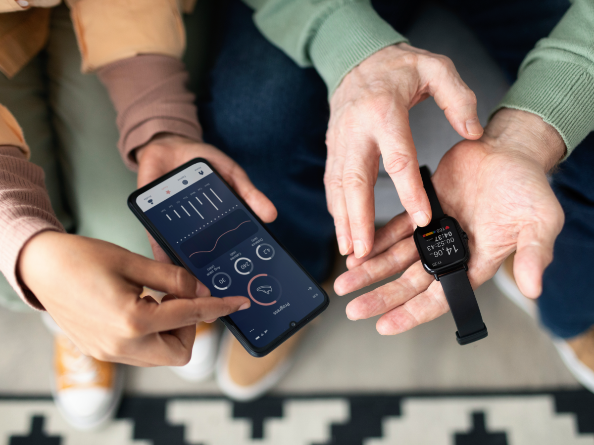
Daily-Life Gait AssessmentsWe measured daily-life gait using in-shoe IMU sensors (A-RROWG, NEC Corporation; ). These sensors are small (40.0 mm × 30.5 mm × 7 mm) and lightweight (11 g), including a 3-axis accelerometer and gyroscope. The IMU sensor in the dedicated insole was placed at the foot arch, and the x-, y-, and z-axes of the IMUs were set along the mediolateral, anteroposterior, and vertical directions, respectively. When a person wearing these sensors walks in a stable straight line over 3 gait cycles between 5 AM and 10 PM, the in-shoe IMU sensor detects that the person is walking based on acceleration in the anteroposterior direction and saves the IMU signals of the next 3 gait cycles as 1 gait measurement []. The IMU signals were sampled at a rate of 100 Hz, transferred to a smartphone via Bluetooth, and stored in a specialized app if participants had one with them. If a person did not have a smartphone, the data were uploaded automatically via Bluetooth at 11 PM by keeping the smartphone near the IMU sensors.
From the saved IMU signals, the mean of 7 gait parameters from 3 gait cycles was calculated and stored on a smartphone, as previously described by Fukushi et al []. The following 7 parameters were calculated:
Gait speed: calculated as stride length (m) divided by stride time (s).Stride length: the distance between the start and end points of the foot trajectory for 1 stride.Dorsiflexion angle: the peak foot angle in the dorsal direction from the ground during the swing phase.Plantarflexion angle: the peak foot angle in the plantar direction from the ground during the swing phase.Foot height: the maximum height of the foot trajectory.Toe-in or toe-out angle: the mean angle of foot adduction or abduction in the direction of the velocity vector during the swing phase.Circumduction: the displacement in the medial-lateral direction during the swing phase.In addition to these 7 gait parameters, the coefficient of variation (CV), calculated as SD divided by mean multiplied by 100, was used to evaluate the variability.
 Figure 1. The in-shoe inertial measurement unit (IMU) sensor. The sensors with accelerometer and gyroscope were set into the dedicated insoles, measured the daily-life gait without the participants’ awareness, and saved the obtained data to their smartphone via Bluetooth. Measurement Protocol
All participants completed a paper-based questionnaire on their general health status, which included fall history in the past year, frequency of stumbling, and fear of falling. Falls at the time of fracture in patients with DRFs were excluded based on the number of falls in the past year.
In the daily gait assessments, we inserted IMU sensors with dedicated insoles into the preferred shoes of both participants’ feet. Each participant was provided a smartphone with only the original app for storing gait data. We verified whether participants could walk using the sensors and whether the sensors worked with a smartphone. Participants were instructed to wear shoes with sensors for 2 to 6 weeks. We requested that participants spend their daily lives as usual; therefore, we did not establish a minimum time for wearing or walking with them. The measurements were recorded without the participants’ awareness, and they could view their most recent gait data by checking their smartphone. In the fracture group, daily gait assessments in the early postoperative period started 2 weeks after DRF surgery to enable the effects of casting or surgery.
To assess baseline functional ability and frailty, HGS [] and the Timed Up and Go (TUG) test [] were performed. HGS was measured in kilograms with a Jamer dynamometer (Sammons Preston). We assessed HGS on the nonfractured side of the fracture group and both sides of the control group. The mean values of 3 measurements were recorded. The time to complete the TUG test was recorded twice: once at the preferred speed and once at the fastest speed. These baseline physical tests were performed 4 weeks after DRF surgery in the fracture group and at the beginning of daily gait measurements in the control group.
To check for long-term functional ability, we asked the fracture group patients to perform the same physical and daily gait assessments again at an outpatient visit 6 months after the surgery.
Data AnalysisThe median and CV of each participant were calculated using the obtained gait data. However, the automatically collected data included hills, turns, and straddling, and we used Smirnov-Grubbs analysis for every gait parameter to exclude any outliers from the data. After exclusion, approximately 20-1000 gait measurements were recorded for each participant during the measurement period, reflecting differences in their lifestyles. Even after the exclusion, gait data included various gait types, such as walking exercise and hurried walking; therefore, we excluded participants with less than 50 gait measurements in either foot, based on a previous report []. Since we computed the data obtained from both feet together, we selected participants with 100 or more gait assessments for this study.
Statistical AnalysisBetween-group differences were assessed using Student t test (2-tailed) for continuous variables in the patient demographics, and the chi-square test for categorical variables. Since the participants’ gait data were not normally distributed, they were assessed using nonparametric (ie, Kruskal-Wallis and Steel) tests. The results of the fracture group in the early postoperative and long-term periods were compared using a paired t test. Statistical significance was set at P<.05.
These analyses were performed using EZR (version 1.55; Division of Haematology, Saitama Medical Center, Jichi Medical University) [].
There were no significant differences in age, body characteristics, or lifestyle variables between the 2 groups. In the section on questions about falls, patients with fractures demonstrated a significantly higher incidence of falls (). None of the fracture group members experienced falls or subsequent fractures after the initial DRF.
Table 1. Participants’ demographics. P values <.05 are considered significant.CharacteristicsControl (n=28)Fracture (n=16)P valueAge (years), mean (SD)62.3 (7.0)65.6 (8.0).20aHeight (cm), mean (SD)155.2 (4.3)154.8 (4.0).77aBody weight (kg), mean (SD)54.4 (8.0)51.6 (8.5).30aBMI (kg/m2), mean (SD)22.6 (3.2)21.5 (3.2).28aHand dominance (right), n (%)27 (96)15 (94).68bFoot dominance (right), n (%)23 (82)14 (88).64bSmoking (current and previous), n (%)5 (18)5 (31).31bAlcohol consumption, n (%)11 (39)5 (31).59bComorbidities, n (%)aIndependent Student t tests were used to compare the groups.
bChi-square test was used for analysis between the groups.
cN/A: not applicable.
Spatiotemporal Data in Daily-Life GaitThe results of the spatiotemporal daily-life gait are presented in . There were no between-group differences in the number of measurements. Compared with the control group, the dorsiflexion angle measured at any time postoperatively in the fracture group tended to be lower and demonstrated no improvement in the postoperative course. Participants in the fracture group demonstrated significant recovery in gait speed, stride length, and plantarflexion angle between 4 weeks and 6 months postoperatively. Among the CV of each gait parameter, only the CV of stride length between the control group and the 4 weeks–postfracture group demonstrated a significant difference.
Table 2. Daily-life spatiotemporal data. P values <.05 are considered significant.CharacteristicsControl group (n=28)Fracture group (n=16)P valueaKruskal-Wallis test was used to compare the control and fracture groups.
bSteel test was used to compare each group.
cPaired sample t test was used for analysis between the groups.
dCV: coefficient of variation.
HGS and Body Balancing AbilityThe HGS in the fracture group demonstrated significant recovery between 4 weeks and 6 months after surgery; however, it was significantly lower in the fracture group compared to the control group. In the TUG test, there were no significant differences between the control and fracture groups or at 4 weeks and 6 months postoperatively in the fracture group ().
Table 3. Physical tests in the control and fracture groups. P values <.05 are considered significant.CharacteristicsControl group (n=28)Fracture group (n=16)P valueaKruskal-Wallis test was used to compare the control and fracture groups.
bSteel test was used to compare each group.
cPaired sample t test was used for analysis between the groups.
dTUG: Timed Up and Go.
We performed daily-life gait assessments with in-shoe IMU sensors and some physical tests, including HGS, to evaluate the differences in spatiotemporal gait and physical ability between patients with DRF and healthy controls. Moreover, we assessed whether these parameters improved during 6 months following DRF. In daily-life gait assessments, patients with DRF demonstrated a lower foot height and higher CV stride length compared to the control group; however, these differences were no longer present 6 months after DRF surgery, reflecting the improvement in some parameters in the fracture group after 6 months of DRF treatment. On the other hand, the dorsiflexion angle in the fracture group tended to be lower consistently during 6 months. In the physical assessments, patients with DRF at 4 weeks and 6 months after the surgery had significantly lower HGS than those without DRF, even though HGS in patients with DRF improved during 6 months.
Some gait parameters, such as gait speed and stride length, improved in the fracture group during 6 months of daily life. Further, the CV of stride length, which is associated with fall risk [], was higher in the fracture group at 4 weeks after DRF and did not significantly change after 6 months. With the development of wearable sensors, spatiotemporal gait characteristics and their variability in daily life have been increasingly identified. However, few studies have explored the long-term changes in gait characteristics related to falls and fractures. As daily-life gait is influenced by various factors, such as environmental and psychiatric factors [], changes in patients with DRF could reflect changes in their lifestyle. Conversely, the risk of subsequent fractures is the highest immediately after the initial fracture []. Although no subsequent fractures occurred in this study, further research is warranted to determine the relationship between gait changes and subsequent fractures. The in-shoe IMU sensor must play an important role in further evaluating this relationship.
The dorsiflexion angle in the fracture group remained lower, whereas other parameters in the fracture group improved during 6 months. As for vertebral fractures, which are typical fragility fractures as well as DRF, patients with symptomatic vertebral fractures walked with shorter and wider strides at the time of injury. Although those with vertebral fractures show improvement in stride time and stride length over time, even reaching healthy levels again, their gait pattern and stability persist for 6 months, implying a greater risk of incident disability among these patients []. Since gait speed is reported to affect other gait parameters, the persistent lower dorsiflexion angle in the fracture group, despite the improvement in gait speed, might be a characteristic of patients with DRF, indicating that patients cannot fully return to healthy states. The dorsiflexion angle in this study, which means the angle between the ground and the sole of the foot, may depend on the movement of all lower extremities. Kyphosis and flexed hip or knee joints, which are common in older people, are related to foot movement or strength []. Although further research is needed to determine the cause of this decrease in dorsiflexion angle, the angle could result in stumbling and falls. Older adults with DRF can be assessed as having a high risk of functional decline, particularly those who have access to a health care facility at an early stage. They should receive appropriate intervention to prevent future falls or fractures along with treatment for the initial fracture. Considering that several previous reports have assessed fall risk using machine learning based on gait data from fallers [,], our results could be effective in creating a more precise machine learning model for evaluating the risk of falls. Further research is warranted to explore not only the cost of developing sensors and apps but also intervention methods and the extent of fall reduction achievable. Nonetheless, our findings using in-shoe IMU sensors outside the hospital could be valuable for future screening tools to evaluate the risk of falls and fractures.
As for physical assessments, the HGS in the fracture group significantly improved during 6 months after DRF, which is consistent with a previous report []. The increased use of the nonaffected hand with DRF in daily life may improve HGS; however, most studies on DRF have focused on the HGS of the affected side, and little is known about that of the nonaffected side. Generally, HGS is associated with health status, including death, falls, and muscle strength []. The improvement in HGS could reflect the improvement in health status, and the lower HGS after 6 months of DRF could be associated with a lower degree of health status in the fracture group. Contrastingly, there were no differences in the TUG test results, contrary to our previous reports [,]. The average TUG test time in both the fracture and control groups in this study was faster compared to previous studies, which may mean that the TUG test, which involves a few steps and seconds in the laboratory, may not reflect the true physical characteristics. Therefore, daily-life gait analysis for a certain period is needed to identify the slight difference between fracture group patients and healthy controls. By using this in-shoe sensor for a certain period, the lower dorsiflexion angle in patients with DRF was revealed, which remained 6 months after the fracture. We would like to further investigate long-term changes in the gait of these patients.
LimitationsThis study had some limitations. First, the number of participants was small, which could have affected the power of this study. However, individual changes over 6 months appeared to confirm the statistical outcomes and might not have affected the overall conclusions of our study. Second, we observed progress up to 6 months after the fractures. The HGS on the affected side continued to improve beyond 1 year. Long-term changes in more patients with DRF should be further explored. Third, the participants were all Japanese, who had the habit of taking off their shoes indoors. Considering that 6 patients with DRF fell inside their houses without shoes, the results may not accurately reflect the daily free-living assessments of barefoot individuals.
ConclusionsIn summary, we performed a case-control study to investigate the long-term changes in HGS and daily-life gait after DRF. Using an in-shoe IMU sensor, we revealed the recovery of spatiotemporal gait characteristics 6 months after DRF surgery without the participants’ awareness. The dorsiflexion angle in the swing phase and HGS were still lower in the fracture group after 6 months, which could be associated with fracture risk. This in-shoe IMU sensor could be useful for evaluating the future fall and fracture risk outside the hospital and for constructing a new health care system related to preventive medicine using wearable devices outside the hospital.
This study was supported by the Japan Society for the Promotion of Science (JSPS) KAKENHI (grant 21K17469) and joint research funding from NEC Corporation. We would like to thank Masahiro Fujimoto, Kanako Nakajima, Shoma Kudo, and Gaku Kutsuzawa for collecting data; Hiroshi Kajitani for collecting data and software development; Akimoto Nimura, Hidetoshi Kaburagi, and Takafumi Koyama for critical analysis and proofreading; and Editage for English language editing.
KF has received joint research funding from NEC Corporation. FN and KN are employees of NEC Corporation.
Edited by L Buis; submitted 05.12.23; peer-reviewed by S Okita, M Nakayama; comments to author 08.01.24; revised version received 29.01.24; accepted 26.02.24; published 20.03.24.
©Akiko Yamamoto, Eriku Yamada, Takuya Ibara, Fumiyuki Nihey, Takuma Inai, Kazuya Tsukamoto, Tomohiko Waki, Toshitaka Yoshii, Yoshiyuki Kobayashi, Kentaro Nakahara, Koji Fujita. Originally published in JMIR mHealth and uHealth (https://mhealth.jmir.org), 20.03.2024.
This is an open-access article distributed under the terms of the Creative Commons Attribution License (https://creativecommons.org/licenses/by/4.0/), which permits unrestricted use, distribution, and reproduction in any medium, provided the original work, first published in JMIR mHealth and uHealth, is properly cited. The complete bibliographic information, a link to the original publication on https://mhealth.jmir.org/, as well as this copyright and license information must be included.
留言 (0)