OBJECTIVES:
To evaluate initial fracture morphology influences on outcomes in simple 2-part pertrochanteric fracture, with a focus on the basicervical component and its initial impaction.
METHODS:
Design:
A retrospective cohort series.
Setting:
Single Level I Trauma Center.
Patients Selection Criteria:
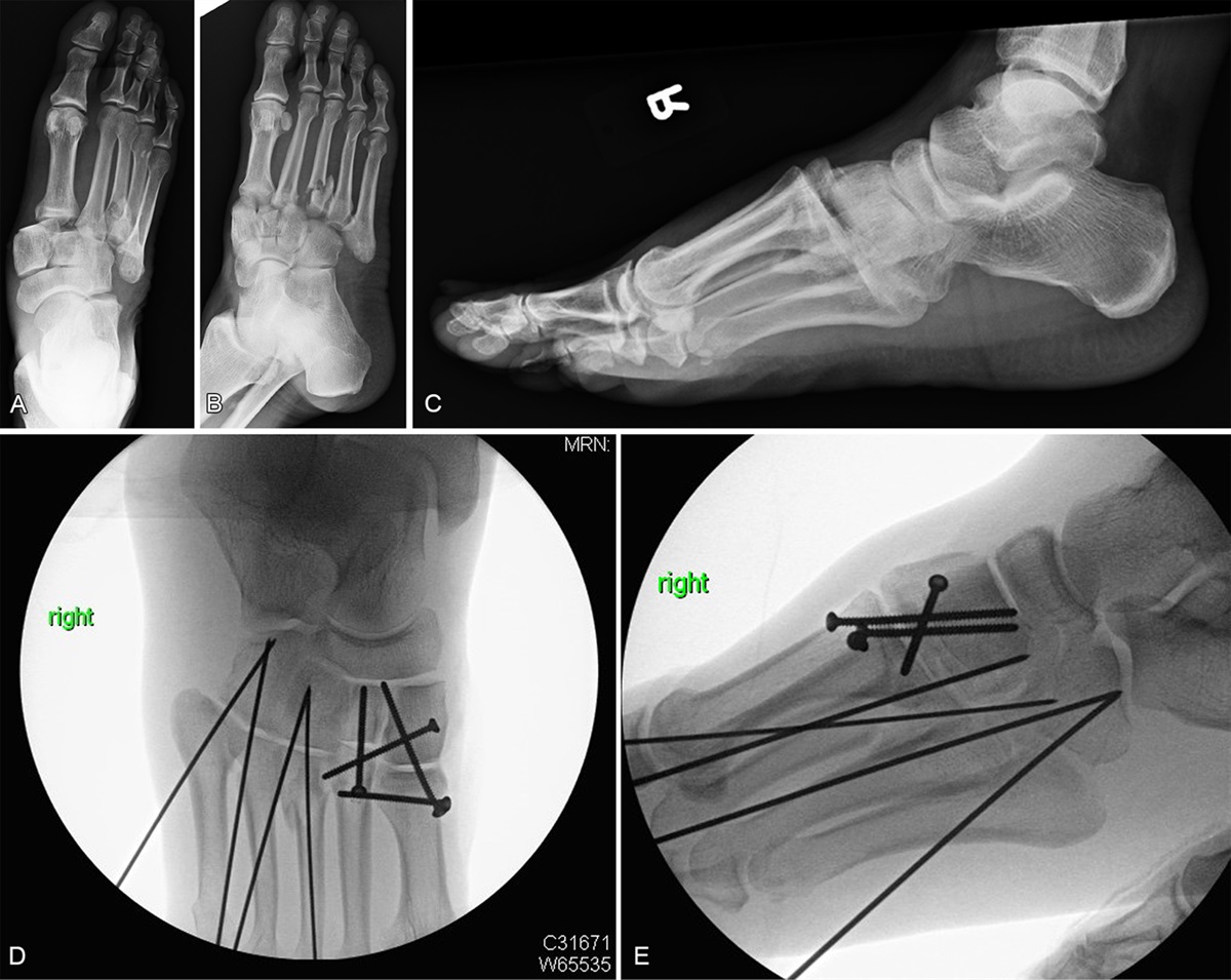
Patients older than 60 years with intertrochanteric fractures between 2011 and 2022 were retrospectively reviewed. Inclusion criteria comprised simple 2-part pertrochanteric fractures (Orthopaedic Trauma Association [OTA]/Arbetisgemeinschaftfur Osteosynthesefragen [AO] 31-A1.2) with a basicervical component who underwent cephalomedullary nailing and had a minimum follow-up of 6 months. Patients were divided whether the basicervical component was impacted into the medullary canal (intramedullary impaction [II] group) or displaced beyond the medullary canal (extramedullary [E] group). Exclusion criteria encompassed pathologic fractures, nondisplaced fractures, and basicervical neck fractures (OTA/AO 31-B3).
Outcome Measurements and Comparisons:
Reduction status was assessed as unacceptable if the head and neck (proximal) fragment was positioned intramedullary with respect to the distal fragment in either the anterior posterior or cross-lateral x-ray and acceptable otherwise. In addition, the degree of impaction on x-ray and CT scans (coronal, sagittal, axial) at injury was analyzed as a risk factor for failure. Revision rates and lag screw sliding over 15 mm were compared between the II and E groups.
RESULTS:
Hundred fifteen patients (95 female, average age 80 years) were included. The II group (n = 58) compared with E group (n = 57) showed more acceptable postoperative reductions (57% vs. 81%, P = 0.001), but significantly higher fixation failure (16% vs. 3.5%, P = 0.048) and fracture collapse (28% vs. 7%, P = 0.01). II was identified as a significant independent predictor for failure (odds ratio 5.64, 95% confidence interval, 2.14–16.9, P < 0.001) with more than 19.5-mm impaction in sagittal CT scan as the threshold linked to increased failure risk.
CONCLUSIONS:
This study highlights the significance of specific intertrochanteric fracture patterns, particularly II of a basicervical component and impaction severity (≥19.5 mm), as drivers of fixation failure.
LEVEL OF EVIDENCE:
Prognostic Level III. See Instructions for Authors for a complete description of levels of evidence.


留言 (0)