
記住我


We present a previously healthy 40-year-old Sri Lankan female who presented with dark urine, malaise, faintness, and body aches for 2 days, and a 1-day history of abdominal cramps and worsening dyspnea without cough or wheeze (Fig. 1). Her previously healthy 8-year-old son was also admitted to the Paediatric services of the hospital with similar symptoms. In both individuals, the symptoms were preceded by a four-day history of self-resolving fever, arthralgia, myalgia, nasal congestion and moist cough. Their family history was not significant up to the point of presentation.
Fig. 1
Dark coloured urine sample suggestive of haemoglobinuria
Upon admission to the general medical ward, the mother had a Glasgow Coma Scale score of 15 (E4V5M6), a pulse rate of 88 beats per minute, and a blood pressure of 140/90 mmHg. She appeared pale, mildly icteric, peripherally cyanosed and afebrile, with no lymphadenopathy or peripheral edema. Although she had a respiratory rate of 20 cycles per minute and no signs of respiratory distress, peripheral pulse oximetry showed alarmingly low levels of oxygen saturation at 70% in room air, which increased to 88% with a nonrebreathing mask providing oxygen at 15 L per minute. Lung auscultation revealed clear breath sounds. Abdominal examination revealed mild, nontender hepatosplenomegaly without free fluid. A point-of-care ultrasound of the lungs, heart, and inferior vena cava to assess fluid status showed normal findings. The mother’s drawn blood appeared dark (Fig. 2). A point-of-care arterial blood gas analysis reported a pH of 7.45 (7.35–7.45), PaO2 of 85.9 mmHg (80–100), SaO2 of 97.9% (> 95), PaCO2 of 23.9 mmHg (35–45), lactate of 0.5 mmol/L (< 2), bicarbonate of 16.9 mmol/L (22–26), base excess of − 7.2 mmol/L, hemoglobin of 8.6 g/dL (11–15), glucose of 110 mg/dL, anion gap of 13.4 mmol/L, PaO2/FiO2 ratio of 410 mmHg, sodium of 135.9 mmol/L (135–145), and potassium of 4.32 mmol/L (3.5–4.5).
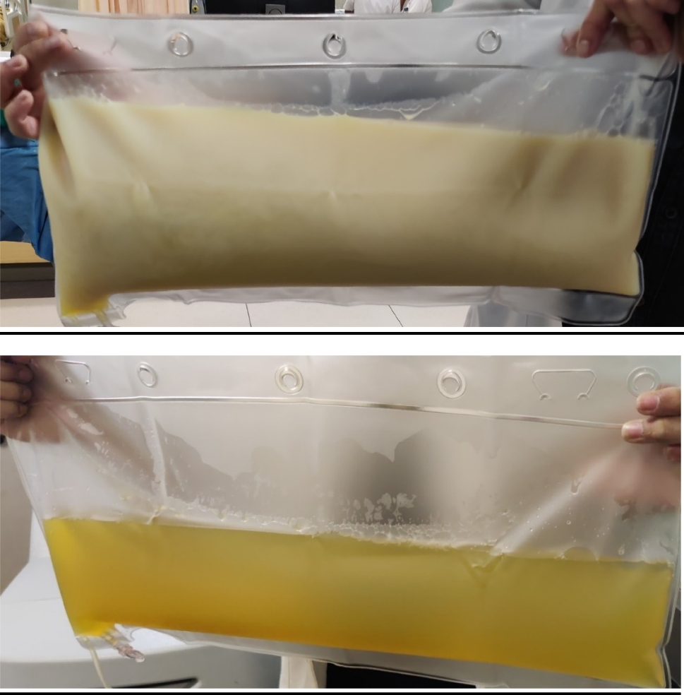
Fig. 2
Dark coloured arterial blood from underlying methaemoglobinaemia
On admission to the paediatric unit, the son was noted to have a normal Body Mass Index (BMI) for age, and normal temperature but to have conjunctival pallor, mild icterus, central and peripheral cyanosis a pulse rate of 100 beats per minute, blood pressure of 100/ 64 mmHg, respiratory rate 32 per minute, and oxygen saturation by peripheral pulse oximetry reading of 65% while breathing room air. The rest of the examination, including the respiratory and abdominal systems, and the fundi was unremarkable. The child also had dark coloured blood and had similar biochemical findings, including a pH of 7.5 (7.35–7.45), PaO2 of 100 mmHg (80–100 mmHg), SaO2 of 98%, PaCO2 of 15 mmHg (35–45), bicarbonate of 20 mmol/L (22–26), haemoglobin of 10.2 g/dL (11–15), sodium of 137 mmol/L (135–145) and potassium of 5.6 mmol/L (3.5–4.5 mmol/L).
Given the initial clinical and biochemical findings of both patients, the possibility of oxidative hemolysis and methaemoglobinaemia was suspected. Upon further inquiry, the mother revealed that she and her son had consumed a popular Sri Lankan herbal remedy, a salad made from shredded leaves of A. indica (Fig. 3) and grated coconut, to help with nasal congestion. The salad had been consumed approximately 12–24 hour prior to the development of dark urine. There was no consumption of any other oxidative medications, such as dapsone or primaquine, fava beans, or exposure to any poisons, such as naphthalene. The child was transferred to the intensive care unit for monitoring.
Fig. 3
Specimen of Acalypha indica
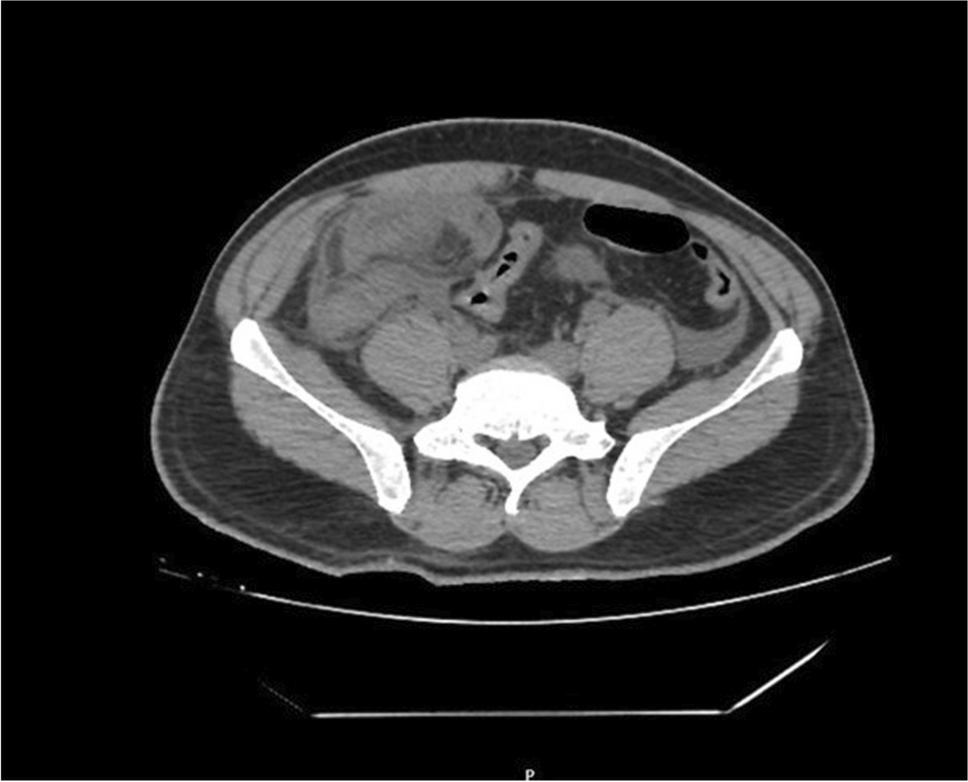
Tables 1 and 2 summarize the trend of investigational findings of the mother and son respectively. An urgent blood film (Fig. 4) of the mother showed evidence of oxidative hemolysis, indicated by numerous spherocytes, polychromasia, nucleated red cells, many blister and bite cells, occasional round and oval macrocytes, and normal platelet count and morphology. Neutrophil leukocytosis with band forms was also noted. Similar findings were observed in the son’s blood picture. The Coomb’s test was negative in both. As G6PD deficiency cannot be confirmed accurately during an acute haemolytic episode, Brewer’s screening test was performed on the only other 13-year-old son, who had not consumed the herb, with the parent’s consent. The test was positive suggestive of an underlying undiagnosed G6PD deficiency in the family. Methaemoglobinaemia was confirmed by spectrophotometry, with levels of 10% detected in the mother and 30% in the child (normal expected value < 2%). Electrocardiogram and chest radiograph of both were normal. Evidence of normal sized hyperechoeic kidneys suggestive of acute renal parenchymal changes was observed in both patients’ abdominal ultrasounds. The mother’s ultrasound also incidentally discovered a coarse hyperechoic liver with splenomegaly of 10.8 cm, consistent with early liver parenchymal disease and portal hypertension.
Table 1 Trend of biochemical investigations of the motherTable 2 Trend of biochemical investigations of the childFig. 4
Mother’s blood picture, bite and blister cells (marked by small arrows) suggestive oxidative haemolysis
In both patients, supportive treatment was initiated, including supplemental oxygen via nonrebreathing mask, blood transfusion (up to 3 packed red cells) to manage anemia, oral folic acid 5 mg daily, oral ascorbic acid 100 mg daily, and conservative fluid management for acute kidney injury. With symptomatic and biochemical improvement of haemolysis and acute kidney injury, both patients were discharged on the 8th day of hospital stay. The parents were educated about the condition, and written information on what herbal remedies, and medications to avoid were provided. At the one-week follow up, both patients had stable haemoglobin, no ongoing haemolysis and return of serum creatinine to baseline. At 4 months post admission, G6PD deficiency in both patients was confirmed using a qualitative G6PD assay. Figure 5 shows the genogram of the patients’ family.
Fig. 5
Genogram illustrating those affected with G6PD deficiency in the family
留言 (0)