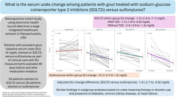
Systemic sclerosis (SSc) is a chronic connective tissue disorder characterized by progressive vasculopathy, autoimmune inflammation, and, eventually, tissue and organ fibrosis [1]. In SSc, calcinosis remains one of the major unmet challenges, as recently addressed in the FDA document [2], thus definitively highlighting the lack of specific treatments. The purpose of our review is to provide an overview of the pathogenesis and management of calcinosis in SSc, highlighting practical approaches and advancements, and identifying current unmet needs.
The National Institutes of Health's National Library of Medicine (PubMed) was searched for publication in English from 01 January 1985 to 01 November 2023 to identify the data. These keywords were used for the screening of publications: (Calcinosis) AND (systemic sclerosis) AND ((pathogenesis) OR (clinical) OR (features) OR (assessment) OR (burden) OR (complications) OR (management) OR (treatment)). The relevant literature including reviews, meta-analyses, original articles, case reports, and series were determined by reading the title and abstract and then these publications were assessed with full text when available to access. In total, one hundred seventy-seven papers were revised.
Calcification of soft tissue is subdivided into five categories, each resulting from distinct underlying etiopathogenic mechanisms: calciphylaxis, idiopathic, metastatic, tumoral, and dystrophic calcification. Dystrophic calcification is defined as soft tissue calcinosis, occurring in patients having normal circulating calcium and phosphorus levels and mostly associated with autoimmune connective diseases [3,4]. In SSc, dystrophic calcinosis is predominantly found in the chemical form of calcium hydroxyapatite [5], [6], [7]. Calcinosis is one of the most common clinical manifestations of SSc, affecting 18 to 49% of the patients, usually occurring in the overexposed areas that are subject to trauma, such as the dorsal parts of the upper limbs, particularly hands, and the knees [8], [9], [10], [11] though occasionally they can occur elsewhere such as the axial spine, paraspinal tissue, temporomandibular joint, and orbital wall [12], [13], [14], [15], [16]. However, the real prevalence and clinical features of calcinosis in SSc patients are yet to be adequately defined. Indeed, calcinosis is often subclinical, that is, found to be much more extensive on imaging (e.g., plain radiography) than determined by clinical examination alone [17,18].
In SSc, the pathogenesis of calcinosis is poorly understood but vascular ischemia and repeated microtraumas are thought to be the key factors driving its development [5,19]. In SSc, progressive vascular damage, including obstructive vasculopathy, is the pathogenetic hallmark resulting in tissue hypoxia and vascular ischemia. At the acral level, clinical studies have demonstrated a strong relationship between calcinosis and severe digital ischaemic manifestations [8,20]. For this reason, chronic ischemia has been suggested as having a role in the pathogenesis of calcinosis, as evidenced by the association of calcinosis with the ulnar arterial occlusion which is detected in around 50% of SSc patients [21,22]. Moreover, loss of digital pulp which is considered a marker of digital ischemia is reported as a risk factor for progression of calcinosis in SSc [23]. However, few studies have demonstrated an increase of hypoxia-related markers in SSc patients with calcinosis. It is now well known that hypoxia plays a central role in the pathogenesis of several metabolic diseases by inducing advanced glycation end products (AGEs) -generated by non-enzymatic glycation and oxidation of proteins or lipids- and the activation of the receptor for AGEs (RAGE) [24]. The AGEs-RAGE interaction leads to vascular damage and calcification by inducing oxidative stress and inflammatory response [25]. Moreover, elevated expression of AGEs and RAGE has been detected in the dermis of limited cutaneous SSc (lcSSc) patients with calcinosis, while elevated circulating levels of RAGE have been reported in SSc patients with digital ischemia [26,27]. A recent study has highlighted that, in the skin of lcSSc patients affected by calcinosis, the expression of glucose transporter molecule-1 (GLUT-1) protein, a marker of cellular hypoxia, is significantly increased compared to lcSSc patients without calcinosis [28]. This evidence has corroborated that hypoxia might contribute to the development of calcinosis in SSc.
In chronic inflammatory and metabolic diseases, the link between inflammation and vascular calcification is a well-known entity [29]. A previous study demonstrated a marked increase in coronary and extra-coronary calcium scores, reflecting vascular calcification, in SSc patients [30]. Among negative acute phase reactant proteins, Fetuin-A has been shown to reduce ectopic calcification by inhibiting mineralization and antagonizing bone morphogenetic proteins [31,32]. In lcSSc patients with calcinosis and diffuse cutaneous SSc (dcSSc), the decrease in serum concentration of fetuin-A suggested its potential use as a novel biomarker for calcinosis [11]. Inorganic pyrophosphate (PPi) has a regulatory role in ectopic calcification by inhibiting the growth of hydroxyapatite formation. A recent study has shown that the serum level of PPi further declined in SSc patients compared to healthy individuals and with no significant difference in circulating PPi levels between patients with and without calcinosis [33].
In SSc, mechanical stress has also been implicated in the development of calcinosis. The observation that the localization and the quantity of calcinosis are most commonly found on the index finger and the thumb may suggest that repeated pinching activity has a role in the genesis of calcinosis [34]. Moreover, an in vitro study has shown that mesenchymal stem cells derived from dcSSc skin have significantly more osteogenic differentiation, an important need for the development of calcinosis, compared to mesenchymal stem cells from healthy participants, in stiff extracellular matrix under stimulation with transforming growth factor beta and interleukin-31 [35]. Overall, this evidence may support the hypothesis that recurrent microtrauma and pressure exposure foster the development of calcinosis in SSc.
From a clinical viewpoint, longer disease duration, older age, anti-centromere, anti-PM/Scl, and anti-RNA polymerase III antibody positivity, a late capillaroscopic pattern, and digital ischemia have been reported to be the major risk factors for SSc-related calcinosis [20,[36], [37], [38]] (Figure 1). A recent study has shown that anti-PM/Scl antibody positivity is significantly related to the burden of calcinosis [39].
Calcinosis represents a great burden for SSc patients due to skin ulceration, infection, fistulation, and consequent disability, which significantly impairs patients’ quality of life. A multi-center prospective study has demonstrated reduced hand function in SSc patients with calcinosis and that impaired hand function is associated with reduced work ability [40]. Moreover, calcinosis is associated with limitation in mobility and predicts the development of disability in SSc patients [8,41]. Furthermore, a study with a small number of patients has demonstrated that the presence of calcinosis is related to reduced quality of life and reported as a negative predictor for the mental component of quality of life evaluated by the Medical Outcomes Short Form-36 (SF-36) [42].
Pain is another significant challenge in SSc, which impacts on the quality of life and work productivity. The majority of SSc patients suffer from pain, and in one-third, the pain is in the moderate or severe range: in particular, SSc patients with calcinosis report a higher degree of pain than those without calcinosis [43]. Therefore, calcinosis can be considered as an independent risk factor for pain [8].
Digital ulcers (DU) are more frequently observed and markedly associated with calcinosis [44,45]. Furthermore, ulcers developed on calcinosis are frequently complicated by infection and fistulation, and in the fingers, DUs associated with calcinosis heal slower than pure (‘ischaemic’) DUs [10,45].
Spinal calcinosis (paraspinal and intraspinal) is not well-known and not easily noticed in SSc patients in contrast to typical dystrophic calcinosis. However, spinal calcification has been reported in a wide range 17 % to 83% of SSc patients, where it may cause neurological symptoms such as numbness, weakness of extremities, and motor and sensory impairments due to spinal cord compression [16,46,47]. Spinal calcinosis has been observed to occur more frequently in male subjects, and patients with DU, and with acro-osteolysis [16]. In addition, paraspinal and intraspinal calcinosis have been reported to be associated with Raynaud's phenomenon, DU, and skin calcinosis [46]. Besides, spinal calcification is detected in more than 80% of SSc patients and pseudo-tumoral calcification is especially located in the intracanal and posterior segments of the spine. The spinal calcification is more frequently observed in SSc patients with periarticular calcification of hands or lcSSc patients [48].
In SSc, patients may complain of severe local pain from calcified masses, which are often called ‘tumoral ’ or ‘pseudo-tumoral’ calcinosis and are usually localized in the spine (especially in the cervical spine). Tumoral calcinosis can also potentially be misdiagnosed as neoplasia or infection and, therefore, needs to be carefully evaluated in SSc patients [13,49,50]. Moreover, tumoral calcinosis when located in the spine can lead to spinal cord compression and even spinal cord injury [51]. Furthermore, a meta-analysis evaluating peripheral neuropathy in SSc has shown that calcinosis is the most frequent risk factor for compressive peripheral neuropathy in SSc patients [52].
A multitude of imaging methods, such as X-ray, computed tomography (CT), magnetic resonance imaging (MRI), and ultrasound (US) can be utilized for the diagnosis, measurement, and quantification of calcinosis in SSc. X-ray is commonly preferred due to its high specificity and sensitivity for the detection of calcinosis as well as its ease of applicability and cost-effectiveness [53]. Besides, an imaging scoring system has been developed using hand radiography, which provides valuable data regarding the severity of calcinosis burden and is employed for follow-up of calcinosis in SSc [23,54]. CT, another useful imaging technique for calcinosis, offers better information about the location, especially when localized in soft tissue, calcinosis-related bone erosions, or associated complications. However, it is important to note that radiation exposure and cost are higher in CT compared to X-ray [51,55,56]. Besides, quantifications of calcinosis can be evaluated with CT using the Agatston score and dual-energy CT applied Convolutional Neural Network [57,58]. Advanced CT modalities such as multidetector imaging or dual-energy CT can be chosen, especially for surgical procedures and treatment follow-up, due to giving detailed data of the calcinosis-located area through enhanced visualization capacities [57,[59], [60], [61]]. The comparison of imaging modalities including X-ray, CT, and MRI in terms of the number and volume/area of calcinosis in hand has elicited that the numbers of detected calcinotic lesions are similar with all three imaging techniques and high correlation has been found between area of calcinosis on X-ray and volume of calcinosis on CT/MRI [62]. US is indeed a highly effective imaging tool, comparable to X-ray, in detecting calcinosis [22,63,64]. Also, US is used as a measurement tool for calcinosis to evaluate the outcomes of treatments [65,66]. A few reported imaging cases have demonstrated extensive calcinosis involvement in the periarticular area and soft tissue by fluorine-18 fluorodeoxyglucose positron emission tomography/CT [67,68].
Considering all of the complications and the burden of calcinosis in SSc patients, the critical question concerns the treatment. Both local or systemic, as well as invasive therapies, have been proposed, but are largely based on anecdotal evidence, due to the absence of standardized outcome measures and randomized controlled studies. There are a few local and various systemic medications that have been used for calcinosis in SSc (Table 1). In calcinosis-related skin ulcers, a study on a small number of patients has evaluated the use of topical Holoil, consisting of neem oil and hypericum perforatum, in addition to standard local treatment (wound care and debridement). Using topical Holoil daily was reported to be significantly associated with improved calcinosis-related ulcer healing (45%), shorter healing time, reduced requirement for antibiotics, and alleviation of pain [69]. Sodium thiosulfate, acting as a calcium chelator, has been used as a local, intralesional, and intravenous systemic form for the treatment of calcinosis, and has also been suggested for SSc-related calcinosis. There are case series reporting the successful use of topical and intralesional sodium thiosulfate, which effectively reduced calcinosis lesions. However, intralesional and topical sodium thiosulfate may cause potential adverse reactions, such as skin irritation, burning sensation, and infection [19,70,71].
The most commonly reported systemictreatments for calcinosis are calcium channel blockers (CCBs), rituximab, minocycline, and colchicine. Overall, there have been conflicting results concerning the efficacy of CCBs on calcinosis in SSc. Case series have reported resolution or regression of calcinosis with diltiazem, whereas a smaller study did not show significant improvement in calcinosis [72], [73], [74], [75]. Similarly, a cohort study suggested that CCBs are protective for calcinosis, whereas longitudinal data did not demonstrate a significant effect on the appearance of new calcinosis [20]. Likewise, a multi-center observational study demonstrated that CCBs did not decrease the risk of calcinosis in SSc patients, except for those with shorter disease duration (<5 years) [76]. As a parallel observation, in studies employing rituximab, a significant remission of calcinosis and clinical improvement has been reported in case reports [77], [78], [79]. Colchicine, a neutrophil tubule inhibitor, may potentially reduce inflammation around calcinosis-related skin ulcers and, therefore, may improve healing, as well as reduce the tissue mineralization, shedding, and inflammatory response of calcinosis [80], [81], [82], [83], [84]. Minocycline, a member of the tetracycline antibiotic family, inhibits poly (ADP-ribose) polymerase (PARP) 1 induced by DNA damage, and overactivation of PARP1 is related to vascular and ectopic calcifications [85,86]. In lcSSc patients with calcinosis, an open-labelled study has shown that 8/9 patients improved calcinosis-related ulceration and infection with minocycline treatment [87]. Considering the association of calcinosis and ischemic findings, the effect of oral Treprostinil, a vasodilator, on calcinosis was evaluated in 12 SSc patients. The results of this study have shown that only 5 patients completed the trial and 80% of these patients had stable calcinosis and 20% with progression of calcinosis [88]. The effect of tofacitinib which is a Janus kinase inhibitor on calcinosis in dermatomyositis is demonstrated by case reports [89,90]. In the literature, only one case report has shown that tofacitinib reduced the extent of calcinosis in refractory scleromyositis [91].
Extracorporeal shock wave lithotripsy (ESWL), mainly employed for the treatment of urinary stone disease, might be an attractive choice for the treatment of calcinosis refractory to surgical and medical therapies [92,93]. A small prospective-controlled study demonstrated that 2/4 patients reported meaningful pain relief, and a reduction in calcinosis was detected in all patients [65]. Another prospective study on 8 patients (3 SSc) also reported the reduction of pain and calcinosis size with ESWL [65].
Surgical intervention is considered for selected patients. For example, surgical excision is the treatment of choice for calcinosis complicated with infection or compressive symptoms. Excision of calcinosis provides attenuation of pain and improves functional impairment [94]. A retrospective study of 39 SSc patients, who underwent surgical debulking of calcinosis in upper extremities, reported a significant decrease in pain and a reduction in the need for opioids. However, only 15% of patients had improvement in their range of motion, likely due to chronic irreversible damage that occurred in surrounding anatomic structures. In the postoperative phase, 44% of patients had complications that were mainly the recurrence of calcinosis, delayed wound healing, and wound infection [95]. In Figure 2, an algorithm for diagnosis and treatment of calcinosis in SSc is provided.


留言 (0)