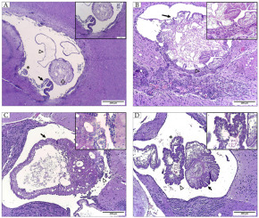
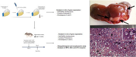
Alveolar echinococcosis (AE) is a serious global zoonosis caused by E. multilocularis and represents a significant disease burden (Casulli, 2020; Dezsényi et al., 2021; Wen et al., 2019). AE is transmitted through the fecal route via exposure to food or water contaminated with E. multilocularis, which is excreted in the feces of foxes or other canines. Domestic dogs are the primary propagators of AE in China (Baumann et al., 2019). AE is characterized by primary tumor-like lesions that induce immunosuppression during chronic infection, a condition also known as parasitic cancer (Brunetti et al., 2010; Calame et al., 2021). E. multilocularis develops predominantly in the liver, mainly in the right lobe, causing lesions with diameters ranging from a few millimeters to >15–20 cm, accompanied occasionally by central necrosis (Eckert et al., 2001; WHO., 1996). Furthermore, unlike E. granulosus, E. multilocularis spreads from the liver to other organs through infiltration or metastasis. Approximately one-third of AE cases are identified incidentally during physical examinations (Eckert et al., 2001; WHO., 1996), although the disease has a high mortality rate, with most patients already exhibiting advanced-stage illness before seeking medical attention (Deplazes et al., 2017; Filippou et al., 2007).
Exosomes are known to participate in the crucial process of intercellular signal transduction in vivo and play a vital role in regulating cell function. Furthermore, their distribution is widespread and stable (Pegtel and Gould, 2019). The latest update in the exosome database (www.exocarta.org) lists 9769 proteins, 3408 mRNAs, 2838 miRNAs, and 1116 lipids (Xie et al., 2019). Exosomes released from parental cells, along with cell-specific cargo, also interact with target cells through receptors on their surfaces and transfer their components to the cytoplasm. This process alters the behavioral and phenotypic characteristics of the target cells, thereby influencing various physiological and pathological processes in these cells, as well as enabling communication with both proximal and distal cells (Nawaz et al., 2019; Zhang et al., 2019).
Exosomes therefore have significant value in both research and clinical efforts to prevent infections by serving as a means of intercellular transport and communication for microorganisms, as well as contributing to immune regulation (Wang et al., 2018). Furthermore, exosomes play a role in antigen/molecule transmission, cellular communication, and cellular immunity, particularly in cases involving parasitic infections within the host (Samoil et al., 2018; Wu et al., 2019). In addition, exosomes derived from parasites contain protected, parasite-specific molecules, including proteins, RNA, ncRNA, miRNA, DNA, and lipids, which are associated closely with parasite virulence and biogenesis (Khosravi et al., 2020).
miRNA is the primary active substance in exosomes and has a regulatory role in host-pathogen interactions through various mechanisms. During parasitic infections, the expression of host miRNAs leads to dysregulation of miRNAs associated with disease progression, resulting in immune and inflammatory responses that contribute to the development of pathogen-associated pathology (Cai et al., 2013; Pockar et al., 2019). Studies have reported that upregulation of specific miRNAs in immune cells during the progression of infection leads to infiltration of immune cells and activation of resident tissue cells, thereby inhibiting transcription processes and inducing inhibition of translation (Cai et al., 2016; Mader and Pantel, 2017).
A substantial amount of evidence indicates that miRNAs, as indispensable cornerstones, hold significant relevance in human parasitic diseases. Based on studies that showed exosomes and miRNAs perform specialized functions in the immune response, this study aimed to characterize these nanoparticles excreted in plasma from AE patients.


留言 (0)