3D anatomy of the supraorbital and greater occipital nerve trajectories
Purpose
This research aims to enhance understanding of the anatomy of the supraorbital nerve (SON) and greater occipital nerve (GON), focusing on their exit points, distal trajectories, and variability, utilizing a novel 3D representation.
Methods
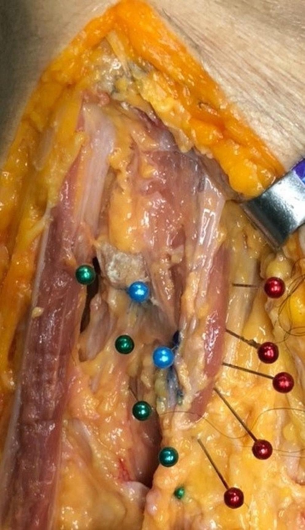
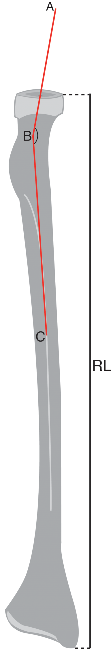
Ten cadaveric specimens underwent meticulous dissection, and 3D landmarks were registered. Models were generated from CT scans, and a custom 3D method was employed to visualize nerve trajectories. Measurements, including lengths and distances, were obtained for the SON and GON.
Results
The SON exhibited varied exit points, with the lateral branches being the longest. The GON showed distinct branching patterns, which are described relative to various anatomical reference points and planes. No systematic left–right differences were observed for either nerve. 3D analysis revealed significant interindividual variability in nerve trajectories. The closest approximation between the SON and GON occurred between lateral branches.
Conclusion
The study introduces a novel 3D methodology for analyzing the SON and GON, highlighting considerable anatomical variation. Understanding this variability is crucial for clinical applications and tools targeting the skull innervation. The findings serve as a valuable reference for future research, emphasizing the necessity for personalized approaches in innervation-related interventions.


留言 (0)