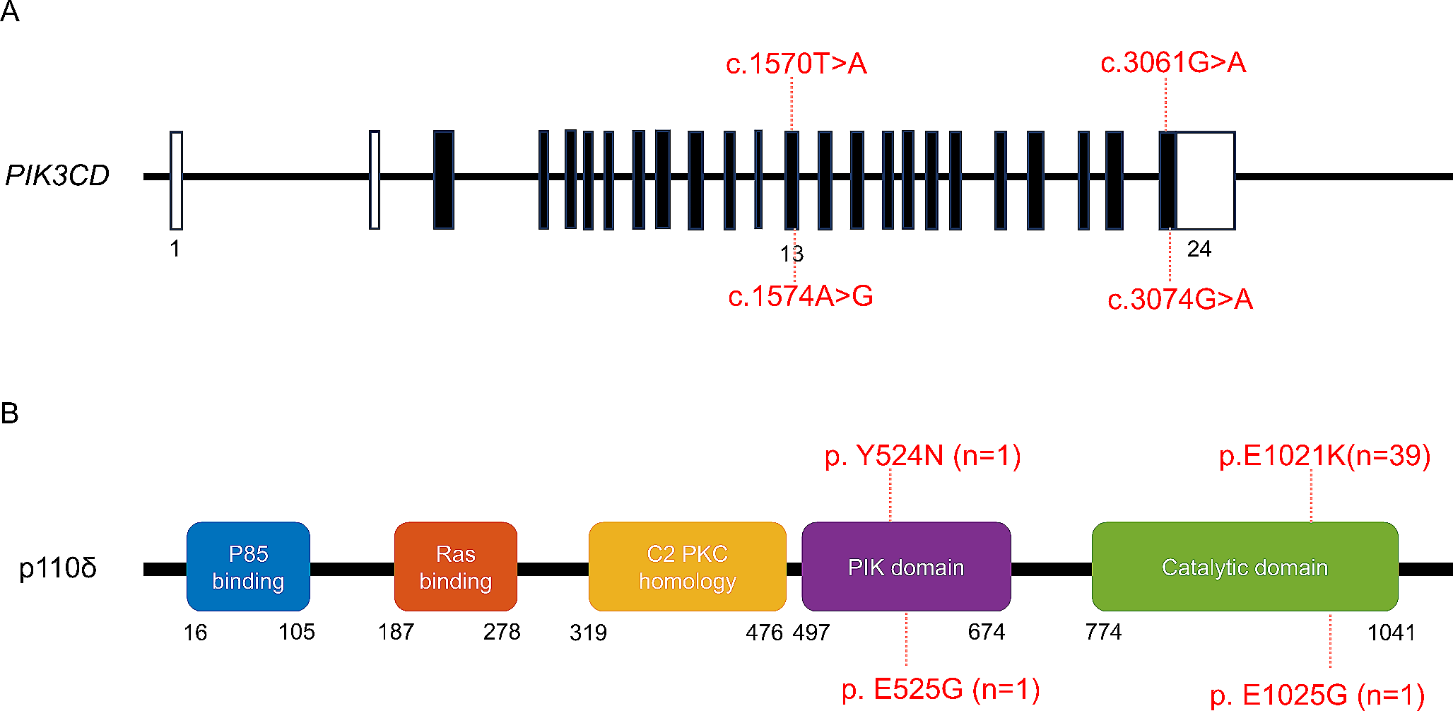
Patient
The patient provided written informed consent to participate in this research in accordance with the Declaration of Helsinki.
Whole Exome Sequencing
Genomic DNA was extracted from the patient’s blood sample and used for whole exome sequencing (WES) at Blueprint Genetics (Helsinki, Finland) (https://blueprintgenetics.com/tests/whole-exome-sequencing).
Immunohistochemistry
Endomyocardial biopsies were collected during myocarditis episode. Samples were fixed in phosphate-buffered 10% formalin (pH 7.0) and embedded in paraffin. The samples were deparaffinized, boiled in Tris-EDTA buffer for 30 min and incubated with Dako REAL Peroxidase-Blocking Solution (Agilent technologies; S2023) for 30 min. The sections were incubated with TIM-3 antibody 1:50 (R&D Systems; MAB23652) for 1-hour at room temperature (RT). After washing, a Dako REAL anti‑rabbit/mouse secondary antibody (Agilent technologies; K500711-2) was added and incubated for 30 min at RT. Target antigens were visualized by using Dako REAL DAB + Chromogen (Agilent technologies; DAKO K500711‐2) for 90 s. Meyer’s Hematoxylin (Sigma-Aldrich; MHS32) was used for counterstaining. Nikon Eclipse 50i microscope equipped with a Nikon DS-Fi3 camera was used for imaging.
Isolation and Culture of Peripheral Blood Mononuclear Cells
Peripheral blood mononuclear cells (PBMCs) from the patient and healthy sex-matched controls were isolated by Ficoll-Paque gradient centrifugation (lithium heparin tubes). The cells were aliquoted in 90% FBS (ThermoScientific; SV301800.03) and 10% dimethyl sulfoxide (DMSO) (Applichem; A3672,0250) and stored at -140 °C. Cells were cultured in RPMI 1640 (Sigma Aldrich; R0883), supplemented with 100U penicillin and 100 µg/ml streptomycin (Sigma Aldrich; P0781), 10mM HEPES (Sigma Aldrich; H0887), 2mM L-glutamine (Sigma Aldrich; G7513) and 10% FBS (ThermoScientific; SV301800.03) at 37℃ with 5% CO2 in a humidified incubator.
Inflammasome Activity Assay
PBMCs from patient and healthy controls were plated at 1.5 × 10⁶/ml density in the conditions described above. The next day, 1 µg/ml lipopolysaccharide (LPS) (Sigma Aldrich; L3012) was applied to the cells for 6 hours followed by an additional 45 min with 5mM ATP (Sigma Aldrich; A6419). The cells were pelleted, and the medium was stored at -80 °C. IL-1β was measured using an enzyme-linked immunosorbent assay (ELISA) (R&D Systems; DY201) according to manufacturer’s instructions. The experiment was performed two times with two healthy controls, all samples were analyzed in duplicates.
Flow Cytometry
In all experiments, PBMCs from the patient and two healthy sex-matched controls were used unless stated otherwise. All flow cytometry samples were analyzed in duplicates unless stated otherwise. The cells were cultured as described above. Phosphate buffered saline (PBS) (Sigma Aldrich; D1408) with 2% FBS (ThermoScientific; SV301800.03) was used for washing, unless stated otherwise. The dead and alive cells were stained with a LIVE/DEAD™ Fixable Near-IR Dead Cell Stain Kit (Invitrogen; L10119) according to manufacturer’s instructions. All antibodies used in the flow cytometry are listed in the supplementary materials. The data was collected with BD LSRFortessa™ using BD FACSDiva software and analyzed with FlowJo™ 10 (BD Biosciences).
Cell Surface and Intracellular Staining of TIM-3 in NK Cells and Monocytes
Freshly isolated PBMCs from patient and healthy controls were plated at 3 × 10⁶/ml density and stained for live and dead cells. For TIM-3 surface staining, the cells were treated with a Fc blocker (BD Biosciences 564,219) for 10 min at RT and for surface markers for 30 min at RT, washed, and fixed for 10 min in 4% formaldehyde (Thermo Scientific; 28,908) at RT. For intracellular TIM-3 staining, the cells were washed after dead and alive staining, permeabilized with Cytofix/Cytoperm (BD Biosciences; 554,714) for 20 min at + 4 °C, washed with cold Perm-Wash (BD Biosciences; 554,714) and treated with a Fc blocker (BD Biosciences 564,219) for 10 min at RT. Then the cells were stained with antibodies for 35 min at + 4 °C.
Cell Surface and Intracellular Staining of TIM-3 in PHA Stimulated T Cells
Freshly isolated PBMCs from the patient and healthy controls were plated at 1 × 10⁶/ml density in supplemented RPMI 1640 medium (2.6), allowed to rest overnight and stimulated with 1.25 µg/ml phytohemagglutinin (PHA) for 4 days. For TIM-3 surface staining, the cells were stained for live and dead cells and surface markers, washed, fixed for 10 min at RT in 4% formaldehyde (Thermo Scientific; 28,908). For intracellular TIM-3 staining, the cells were stained for live and dead cells, washed, permeabilized with Cytofix/Cytoperm (BD Biosciences; 554,714) for 20 min at + 4 °C and washed with cold Perm-Wash (BD Biosciences; 554,714). Finally, the antibodies were added and incubated for 35 min at + 4 °C.The intracellular TIM-3 median fluorescence intensity (MFI) value was calculated with the following formula: MFI TIM-3 obtained from intracellular staining – MFI TIM-3 obtained from surface staining. The TIM-3 expression was first analyzed on the surface of PHA induced CD4+ and CD8+ T lymphoblasts and further confirmed on CD56+ natural killer (NK) cells and CD14+ monocytes. All experiments included two healthy controls and all procedures were done in duplicates.
Expression of Checkpoint Inhibitor Receptors LAG-3, TIM-3 and PD-1
PBMCs from patient and healthy controls were plated at 2.5 × 10⁵/ml density in supplemented RPMI 1640 medium (2.6) and allowed to rest overnight. The cells were stimulated with 8 µg/ml Phytohemagglutinin (PHA-P) (Sigma Aldrich; L-1668) for 3 days. The cells were washed and stained for live and dead cells and surface markers for 30 min at RT, washed and fixed with 4% formaldehyde (Thermo Scientific; 28,908). The experiment was performed once with two healthy controls, all stained samples were done in duplicates.
Regulatory T Cell Analysis
PBMCs from patient and healthy controls were plated at 3 × 10⁶/ml in supplemented RPMI 1640 medium (2.6). The cells were allowed to rest overnight and stained for live and dead cells. The cells were stained for surface markers in Brilliant Stain Buffer (BD Biosciences; 563,794) for 30 min at 4 °C, washed, fixed and permeabilized with Transcription Factor Staining Buffer Set (Miltenyi Biotec; 130-122-981) for 30 min at 4 °C. After permeabilization the cells were washed and stained for FOXP3 for 35 min at 4 °C. The experiment was performed twice with two and four healthy controls, all stained samples were done in duplicates.
T Cell Proliferation
PBMCs from patient and healthy controls were plated at 3 × 10⁶/ml density in supplemented RPMI 1640 medium (2.6), allowed to rest overnight, washed and stained with CellTrace™ CFSE Cell Proliferation Kit (Invitrogen; C34554) for 8 min at + 37 °C. Cold supplemented RPMI was added and incubated for 10 min at RT. Then the cells were centrifuged and resuspended in RPMI medium supplemented as described above (2.6). The cells were plated at 1 × 10⁶/ml and following stimulants were added: 2.5 µg/ml or 1.25 µg/ml PHA-L solution (Invitrogen; 00-4977-93) or Anti-CD3/anti-CD28 beads (Gibco; 11131D), CD3 (Miltenyi Biotec; 130-093-387) and CD28 (Miltenyi Biotec; 130-093-375) antibodies, all stimulants were done in triplicates both with or without 100U/ml IL-2 (Peprotech; #200-02). The cells were stimulated for 4 days and stained for CD4 and CD8 surface markers before acquisition. The experiment was performed twice with two healthy controls, all stained samples were done in triplicates.
Intracellular Staining of Interleukin 2 (IL-2)
PBMCs from patient and healthy controls were plated 3 × 10⁶/ml density in supplemented RPMI 1640 medium (2.6) and allowed to rest overnight. A protein transport inhibitor containing Brefeldin A and Monensin (Invitrogen; 00-4980) was added to unstimulated cells or the cells were stimulated for 5 h with PMA/Cell Stimulation Cocktail with protein transport inhibitors (Invitrogen; 00-4975-93). The cells were collected, washed, and stained for live and dead cells and surface markers for 30 min at RT. The cells were washed, permeabilized with Cytofix/Cytoperm (BD Biosciences; 554,714) for 20 min at + 4 °C, washed with cold Perm-Wash (BD Biosciences; 554,714) and stained for 35 min at + 4 °C with IL-2 antibody. The experiment was performed twice with two healthy controls, all stained samples were done in duplicates.
Intracellular IFN-γ Staining
PBMCs from patient and healthy controls were plated at 3 × 10⁶/ml density in supplemented RPMI 1640 medium (2.6) and allowed to rest overnight. Protein transport inhibitors (Invitrogen; 00-4980) were added to unstimulated cells, or the cells were stimulated for 5 h with PMA/Cell Stimulation Cocktail with protein transport inhibitors (Invitrogen; 00-4975-93). After stimulation, the cells were collected, washed, and stained for live and dead cells and surface markers. The cells were washed twice, permeabilized with Cytofix/Cytoperm (BD Biosciences; 554,714) for 20 min at + 4 °C, washed twice with cold Perm-Wash (BD Biosciences; 554,714) and stained with IFN-γ antibody for 35 min at + 4 °C. The experiment was performed twice with two healthy controls, all stained samples were done in duplicates.
STAT Phosphorylation
For signal transducer and activator of transcription (STAT) 1 and 3 previously described methods were used [16, 17]. For STAT4, PBMCs from patient and healthy controls were plated at 4 × 10⁶/ml density in supplemented RPMI 1640 medium (2.6) and allowed to rest overnight. The cells were subjected to a 3-day pre-stimulation with 1.25 µg/ml PHA-L (Invitrogen; 00-4977) and 100U/ml IL-2 (Peprotech; #200-02), collected, washed, and stained for live and dead cells. The cells were washed, resuspended in medium and allowed to rest for one hour. The cells were stimulated with 10ng/ml recombinant IL-12 (Peprotech; #200 − 12) for 20 min at 37 °C and immediately fixed 10 min in 4% formaldehyde (Thermo Scientific; 28,908) at RT. After two washes, the cells were permeabilized with ice cold Perm Buffer III (BD Biosciences; 558,050) for 30 min on ice, washed and stained for 35 min at + 4 °C with antibodies. The STAT4 experiment was performed twice with two healthy controls, all stained samples were done in duplicates.


留言 (0)