Shareable abstract
The PIOPED II study provided a robust estimate of the diagnostic accuracy of multidetector CTPA in suspected pulmonary embolism and played a pivotal role in establishing CTPA as the current diagnostic gold standard https://bit.ly/3HEyVxy
Introduction
Pulmonary embolism occurs when one or more branches of the pulmonary arterial vessels are obstructed by material originating from elsewhere in the vasculature. Diagnosing this condition may be complex because of nonspecific symptoms that vary based on the extent of pulmonary vasculature obstruction and individual patient characteristics. Typically, patients experience some degree of dyspnoea or chest pain, creating a clinical presentation similar to other emergency conditions such as acute coronary syndrome, pneumothorax, or heart failure.
The recommended diagnostic approach varies little across medical societies [1]: a clinical assessment that does not provide an alternative explanation for the patient's symptoms should be followed by an evaluation of the pretest probability of pulmonary embolism, for instance by using the Wells' criteria. Subsequently, in cases of low to intermediate pretest probability D-dimer levels are measured. Elevated D-dimer levels or a high pretest probability prompt further investigation with computed tomography pulmonary angiography (CTPA). However, this nearly universal consensus has not always been the norm.
Approach to the diagnosis of pulmonary embolism in the pre-CTPA era
In the past, when clinicians suspected pulmonary embolism they relied on chest radiographs, electrocardiograms and arterial blood gases. However, these methods often provided insufficient information, leaving clinicians heavily dependent on their clinical assessments. This posed a dilemma: deciding between initiating anticoagulation therapy, which carried the risk of haemorrhage, or refraining from treatment, which could increase the risk of recurrence and mortality.
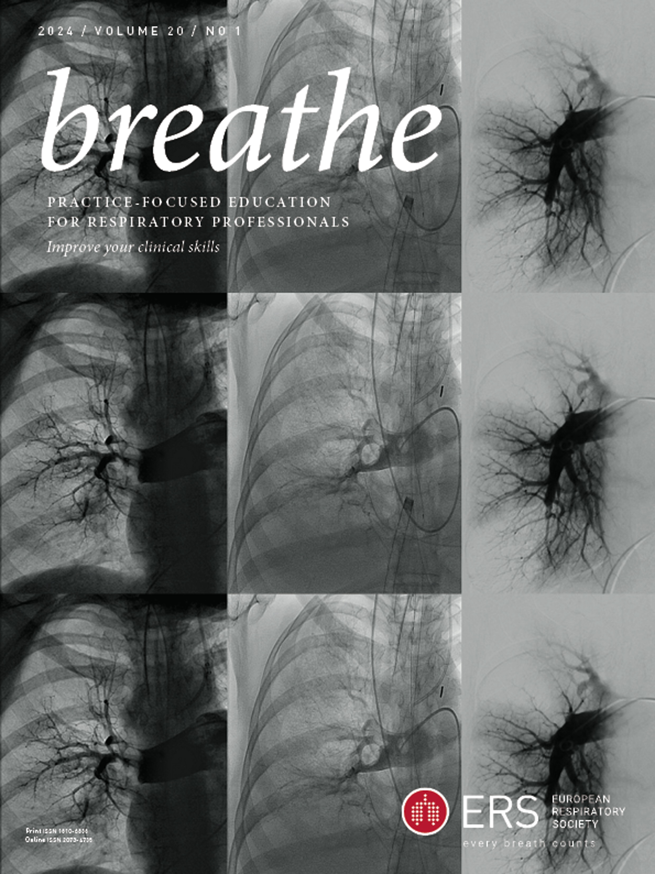
In the mid-twentieth century, the introduction of pulmonary angiography marked a significant advancement. This invasive procedure involves injecting iodinated contrast agent directly into the pulmonary arteries under fluoroscopic guidance, through a catheter inserted in a femoral vein. A thrombus manifests as a filling defect or an abrupt cessation of blood flow, confirming the presence of pulmonary embolism. Use of pulmonary angiography was limited to specialised centres with trained operators and carried a mortality rate of up to 0.3% [2]. Moreover, its interpretation is challenging and prone to underestimating segmental defects.
In 1968 and 1970, Wagner et al. [3] and DeNardo et al. [4] proposed an alternative method for pulmonary embolism diagnosis with combined ventilation and perfusion (Vʹ/Qʹ) lung scintigraphy. A characteristic image of pulmonary embolism would appear as a Vʹ/Qʹ mismatch, indicating areas of pulmonary vasculature obstruction with preserved ventilation. Nevertheless, interpreting Vʹ/Qʹ scan images can be difficult, especially in patients with underlying cardiopulmonary diseases, and it was not until 20 years later that the Vʹ/Qʹ scan became a universally accepted approach for suspected pulmonary embolism.
In 1990, the PIOPED (Prospective Investigation of Pulmonary Embolism Diagnosis) study compared the efficacy of Vʹ/Qʹ scintigraphy to that of pulmonary angiography as the reference standard [5]. The study underscored the limitations of Vʹ/Qʹ scintigraphy, revealing a low sensitivity rate and its failure to identify 59% of patients with acute pulmonary embolism. Most of these undetected cases involved scans showing intermediate or low probability, making conclusive diagnoses challenging. Subsequent retrospective analysis of the PIOPED scintigraphic criteria and additional prospective studies demonstrated that modifying the original reading criteria enhances test performance [6–8]. While these revised PIOPED diagnostic criteria are still recommended to this day, Vʹ/Qʹ scintigraphy did eventually get challenged as the test of choice for suspected pulmonary embolism [9].
Design and results of the PIOPED II-study
The PIOPED II study was a pioneering study within research on the diagnostic work-up of acute pulmonary embolism [10]. It was published under the title “Multidetector computed tomography for acute pulmonary embolism” by Stein et al. [10] in the New England Journal of Medicine in 2006.
The study was sponsored by the National Heart, Lung, and Blood Institute and was conducted as a prospective multicentre study across eight clinical sites in North America between September 2001 and July 2003. The aims were: 1) to determine whether multidetector CTPA could reliably detect and rule out acute pulmonary embolism; 2) to explore whether the addition of venous phase multidetector computed tomography (CT) venography (from the inferior vena cava confluence through the popliteal veins) improved diagnostic accuracy; and 3) to investigate whether the addition of a clinical assessment (Wells' score) improved the clinical utility of CTPA with and without the addition of CT venography.
A total of 7284 patients ≥18 years old suspected of acute pulmonary embolism were screened for inclusion. Of these, 3262 were eligible for inclusion, 1090 patients were enrolled, and 824 patients received both CTPA and CT venography as well as one or more of the components which together formed the composite reference standard. The components included Vʹ/Qʹ scintigraphy, doppler ultrasonography of the lower extremity veins, and if necessary digital subtraction pulmonary angiography. The assessments of all images were performed by two blinded radiologists unaffiliated with the study institutions.
Among these 824 patients, 192 (23%) were diagnosed with acute pulmonary embolism according to the composite reference tests. For those with sufficient diagnostic image quality (n=773 patients), CTPA alone had a sensitivity of 83% (150 of 181 patients) and a specificity of 96% (567 of 592 patients). The positive predictive value was 86% (150 of 175 patients) and the negative predictive value was 95%. Combined CTPA and CT venography, in patients with sufficient image quality, increased the sensitivity to 90% without affecting specificity. The positive predictive value was 85% (164 of 194 patients) and the negative predictive value was 97% (524 of 543 patients) (table 1).
TABLE 1
The main results of the PIOPED (Prospective Investigation of Pulmonary Embolism Diagnosis) II study
Predictive values of CTPA varied substantially by clinical suspicion, with a positive predictive value of 96% among those with a high pre-test probability of acute pulmonary embolism and of 58% among those with a low pretest probability (table 1).
In conclusion, the study found that multidetector CTPA solely, or potentially in addition to CT venography, had a high sensitivity and specificity for acute pulmonary embolism, making it a valuable tool in the diagnosis of this condition.
Diverging perspectives: PIOPED II's legacy and the CT venography conundrum
The reception of the PIOPED II study within academic circles was generally positive, albeit tinged with nuances among key researchers in pulmonary embolism diagnosis. A 2006 editorial in the New England Journal of Medicine by Perrier and Bounameaux [11], pioneers in D-dimer usage for suspected pulmonary embolism, declared that the study convincingly established the diagnostic value of multidetector CTPA. However, they cautioned against the routine use of CT venography, asserting that it didn't sufficiently enhance the diagnostic yield to justify the additional radiation exposure [11]. This perspective diverged notably from that of the PIOPED II authors, who underscored the superior sensitivity (90%) of combined multidetector CTPA and CT venography over CTPA alone (83%) [10], and recommended the use of both CTPA and CT venography, irrespective of pre-test probability, in their 2006 diagnostic algorithm [12]. When considering this discrepancy in the interpretation of the PIOPED II study data, it should be noted that Perrier et al. [13] had published a study a year earlier, reporting a promising 3-month failure rate of 1.5% (95% CI 0.8–3.0) in 756 patients where a D-dimer assay and multidetector-row CT angiography were the sole tests used to rule out pulmonary embolism.
The Fleischner Society, an international society for thoracic radiology, also took notice of the findings of PIOPED II [14]. In its 2007 statement on management of suspected acute pulmonary embolism, it was acknowledged that the PIOPED II data supported the use of multidetector CTPA as a first-line examination for suspected pulmonary embolism, but not as a stand-alone imaging technique. However, when taking into account the study by Perrier et al. [13] as well as the Christopher study [15], in which pulmonary embolism was also ruled out by CTPA and D-dimer alone in 1028 patients with a 3-month failure rate of 0.5% (95% CI 0.2 to 1.1), the Fleischner Society recommended the addition of CT venography only in selected cases on the basis of risk–benefit considerations [14]. In particular, the recommendations advised to avoid CT venography when evaluation of the lower extremity veins was not important clinically or in female patients of reproductive age [13–15].
What then emerged as the favoured diagnostic approach? CTPA alone or in tandem with CT venography? In the aftermath of the PIOPED II study's recommendations, 11 guidelines on pulmonary embolism diagnosis were issued by expert groups and medical societies [9, 16–25]. With the exception of the European Association of Nuclear Medicine, all guidelines advocated for CTPA alone as the primary modality [1].
While the recommendations from the PIOPED II study were not universally implemented in subsequent guidelines, probably due to CT technical improvement allowing higher image quality and increased sensitivity for pulmonary embolism detection, the study results continue to play a pivotal role in contemporary academic discussions on CTPA. PIOPED II stands out as the only study among those mentioned that subjected all participants to both CTPA and a reference standard, enabling accurate estimation of sensitivity and specificity. The reported sensitivity of 83% and specificity of 96% in PIOPED II are still considered among the best estimates today, referenced by, for instance, the 2019 collaborative guidelines of the European Respiratory Society and European Society of Cardiology [9].
These values as likely to increase in the coming years, as wider availability of artificial intelligence software may enhance radiologists' sensitivity in detecting pulmonary emboli, and photon-counting detectors may further enhance CTPA performance while lowering contrast-medium volume and radiation dose [26, 27].
Summary
The PIOPED II study provided a robust estimate of the diagnostic accuracy of multidetector CTPA in suspected pulmonary embolism and the results remain the mainstay in the contemporary academic discussion on the topic. While the authors' recommendation that CTPA and CT venography should be used in tandem to increase sensitivity was not adopted by future societal guidelines, the study played a pivotal role in establishing CTPA as the current gold standard in diagnosis of pulmonary embolism.
Footnotes
Conflict of interest: P.I. Pietersen has received a travel grant from Boehringer Ingelheim. C. Goyard has no conflicts of interest to declare. T. Gille reports personal fees from Roche S.A.S., and other support from Oxyvie (oxygen provider), Vivisol France (oxygen provider), and Menaniri France outside the submitted work. C. de Margerie-Mellon has received fees from Bracco outside the submitted work. C. Falster has received personal fees from Bristol-Myers Squibb and AstraZeneca outside the submitted work.
Received November 28, 2023.Accepted January 25, 2024.
http://creativecommons.org/licenses/by-nc/4.0/Breathe articles are open access and distributed under the terms of the Creative Commons Attribution Non-Commercial Licence 4.0. For commercial reproduction rights and permissions contact permissionsersnet.org


留言 (0)