Acute necrotizing duodenitis in diabetic ketoacidosis
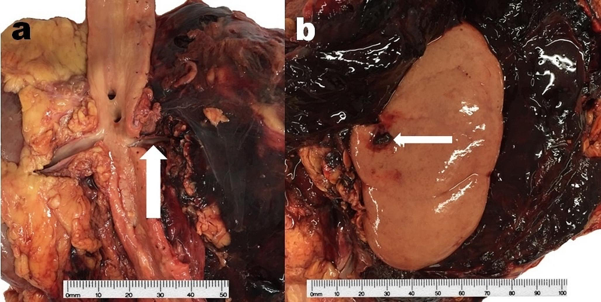
This case documented isolated AND (or ‘black duodenum’), in a death from pneumonia complicated by DKA. The peculiar features in this case are not only the isolated presentation but also its overlapping morphology with other upper gastrointestinal lesions associated with DKA, namely acute necrotizing esophagitis (ANE or ‘black esophagus’) and Wishnesky’s lesions (WLs). This case provides insight into the pathogenesis of these peculiar gastrointestinal findings, and documents the variation in presentation of AND in the context of DKA.
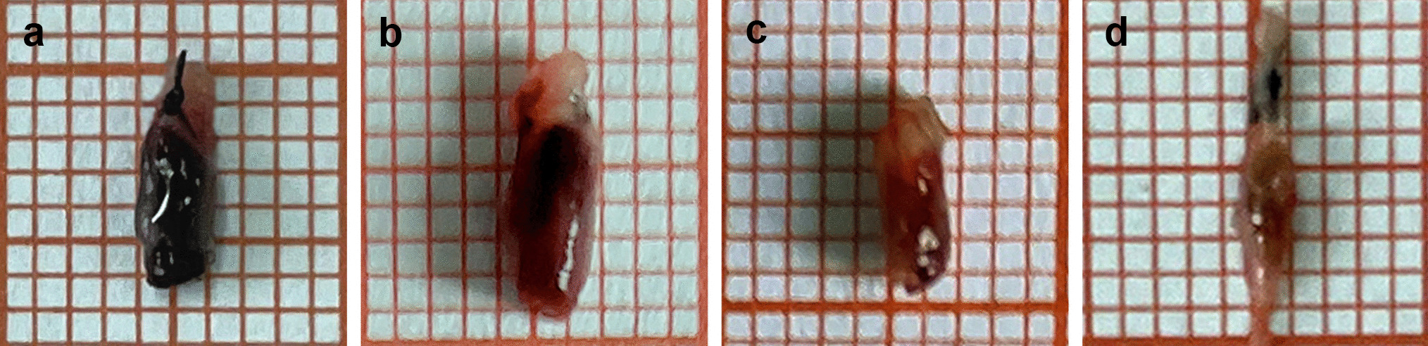
Literature described the different morphological appearances between ANE, WLs and AND in their respective locations. Proposed overlapping mechanisms that cause these lesions include general metabolic derangement and local factors such as blood perfusion, and exposure to gastric content (gastric acid and enzymes) [1,2,3,4]. Macroscopically, ANE presents as generalised black discolouration of the oesophagus propagating from the distal oesophagus and extending proximally and has a definite demarcation at the gastro-oesophageal junction [4, 5]. WLs are black lesions ranging from punctate dots to up to 40 mm on the gastric mucosa [3, 6]. AND is recently reported in deaths from DKA in two separate case reports (with photograph) and one from sepsis (without photograph) in the forensic pathology literature [1, 7, 8]. The two case reports related to DKA described/documented AND to have the macroscopic appearance resembling ANE with either generalised black discolouration or a singular dark lesion on the duodenum which have a definite demarcation at the gastro-duodenal junction [1, 6, 8, 9]. Furthermore, the AND in both cases were seen with ANE and WLs. In the presented case, the AND was isolated without ANE and WLs, and macroscopically it was discrete and patchy resembling WLs, which differed from what was previously described.
Histologically all three conditions have dark granular collections and necrotic features. Different from WLs, ANE and AND have pronounced neutrophilic and lymphocytic infiltration of the mucosa [1, 6, 9]. Despite the morphology of the duodenal lesions in this case resembling WLs, the histology resembles that of AND in keeping with previous case reports. A summary of the macroscopic and microscopic morphology of these three lesions is shown in Table 1.
Table 1 Summary of morphological and histological features of three gastrointestinal features found in diabetic ketoacidosis: acute necrotising esophagitis, Wishnesky’s lesions, and acute necrotising duodenitis


留言 (0)