Trichomonas vaginalis is an extracellular flagellate protozoan and the etiological agent of human trichomoniasis, a sexually transmitted infection (STI) with a high incidence. It is now well recognized that most, if not all, cells from very different organisms secrete macromolecules and vesicles into the extracellular environment. These are generally designated as extracellular vesicles (EVs), which can be secreted spontaneously and stimulated under certain conditions, such as increased calcium concentration (Reviews in Chitti et al., 2024; De Souza and Barrias, 2020; Raposo and Stoorvogel, 2013; Sedgwick and D'Souza-Schorey, 2018). They may have diameters varying from approximately 30 nm to a few micrometers and have been found in body fluids such as blood, urine, cerebrospinal fluid, breast milk, and saliva, as well as in culture media (Review in Sedgwick and D'Souza-Schrey, 2018). They have also been associated with different biological processes in eukaryotic and prokaryotic cells (Sedgìck and D'Souza-Schorey, 2018; Raimondo et al., 2011; Raposo and Stoorvogel, 2013; Kim et al., 2015; Yáñez-Mó et al., 2015; Théry et al., 2018; Monguió-Tortajada et al., 2019).
Extracellular vesicles (EVs) shed by eukaryotic cells into the extracellular space can be divided into subgroups. Exosomes (E) originate from multivesicular bodies (MVB), a component of the endocytic pathway, while ectosomes (EC) are formed from a budding process on the cell surface and present a diameter varying between 100 and 1000 nm (Kang et al., 2021; Yorgy et al., 2011; Reviews in Camussi et al., 2010, De Souza and Barrias, 2020, Raposo and Stoorvogel, 2013, Tkach and Théry, 2016, Yáñez-Mó et al., 2015). Apoptotic or dying cells also release vesicles with a 1000–5000 nm diameter. In addition, other processes, including oncosomes, migrasomes, and exospheres, have also been reported (Review in Chitti et al., 2024). In addition, vesicles are released from the cell surface after a patching and plugging process of ligand/receptor complexes. The last subgroup involves other processes in which tubules are secreted by a parasite that produces structures like exosomes (Review in De Souza and Barrias, 2020).
Microvesicles are important in cell-to-cell communication, tissue homeostasis, cell differentiation, and organ development and remodeling (Tkach and Théry, 2016). Studies on mammalian cells indicate that during the process of budding and release of microvesicles, there is an increase in the concentration of cytoplasmic calcium (Ca2+) and a loss of asymmetry of phospholipids in the plasma membrane (Taylor et al., 2020). Elevated intracellular Ca2+ is required in microvesicle biogenesis and secretion. Still, despite its major role in this process, the source of Ca2+ mobilization (intra- or extracellular) leading to plasma membrane vesiculation remains elusive (Taylor et al., 2020).
Parasitic protozoa also secrete EVs considered exosomes and ectosomes (Reviews in De Souza and Barrias, 2020; Wang et al., 2022). Several reports have shown that T. vaginalis releases EVs into the culture medium, which shows high potential in modulating cell-to-cell communication and the host response to infections (Twu et al., 2013; Nievas et al., 2018; Olmos-Ortiz et al., 2017). This protozoan releases microvesicles of variable size after specific physiological stimuli (Nievas et al., 2018).
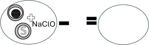
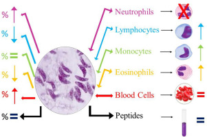
Initially, T. vaginalis EVs were characterized as exosomes based on the description of the ultrastructure and proteomic analysis (Twu et al., 2013). Scanning electron microscopy revealed the presence of cell surface protrusions, which may correspond to sites of vesicle release in around 1% of untreated cells, increasing to around 10% after incubation for 30 min in the presence of 1 mM calcium chloride (Nievas et al., 2018). Exosomes secreted by T. vaginalis demonstrate an important immunomodulatory role in the profile of cytokines induced by the parasite in the expression of interleukin-10 (IL-10), IL-6, and TNF-α (Olmos-Ortiz et al., 2017). Twu et al. (2013) showed that exosomes are released and modulate host-cell immune responses. The study of these vesicles is challenging due to the complexity of their biogenesis and the vast heterogeneity of size, composition, and origin when they are already released. Thus, it was necessary to develop techniques to detect and characterize them. Some are based on biophysical properties and molecular and fluorescent markers. The most used detection procedures are western blotting, nanoscale tracking analysis, and electron microscopy (Aimaletdinov and Gomzikova, 2022; Lakkaraju and Rodrigues-Bouln, 2008; Raimondo et al., 2011; Silverman et al., 2010; Thery et al., 2018 Van der pol et al., 2010).
EVs may carry Rab GTPases or proteins involved in the stimulus of the formation of multivesicular bodies (Raposo and Stoorvogel, 2013; Théry et al., 2009; Taylor and Gercel-Taylor, 2011), conserved proteins such as heat shock proteins (HSP60, HSP70, HSP90), proteins with adhesion activity (tetraspanins, CD81, CD63, CD37), annexins (I, II, V, VI), cytoskeletal proteins (actin, tubulin), metabolic enzymes, and proteins with translational activity (elongation factors) or signaling (Raposo and Stoorvogel, 2013; Théry et al., 2009; Taylor and Gercel-Taylor, 2011; Schorey and Bhatnagar, 2008; Simpson et al., 2008; Silverman et al., 2010; Yang and Robbins, 2011; Atayde et al., 2015). In addition, RNAs found in these extracellular vesicles include several biotypes representing a selected portion of the RNA content of the cell of origin, with a strong tendency to comprise small non-coding RNAs (O'Brien et al., 2020).
The RNA cargo of T. vaginalis vesicles is rapidly internalized by human cells through lipid raft-dependent endocytosis. This payload predominantly comprises tRNA-derived small RNAs (tsRNAs), an emerging class of small regulatory RNAs. It requires further studies and suggests a new mode of host-cell communication in this parasite (Olmos-Ortiz et al., 2017). Exosomes seem to mediate both host-parasite and parasite: parasite interactions. In addition, they play a role in the attachment of T. vaginalis to host epithelial cells (Twu et al., 2013). T. vaginalis vesicles cannot only deliver their soluble content but also transfer lipids when the exosome membrane fuses with the membrane of host cells (Artuyants et al., 2020). The general mechanisms driving vesicle uptake and cargo delivery have not been fully characterized. The heparan sulfate molecules in host cells seem to act as receptors in the first step of internalizing these vesicles (Rai and Johnson, 2019).
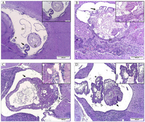
Despite all the basic information on EVs secreted by T. vaginalis, there is little information on the biogenesis of the vesicles, their secretion at the protozoan surface, and their general shape. Therefore, we decided to conduct further morphological analysis to answer some questions not addressed in previous studies (Nievas et al., 2018). These include (a) analysis of vesicle release and surface morphology at shorter incubation times, (b) observations at a much higher resolution using high-resolution scanning microscopy, including electrons and Helium ions, (c) analysis of the effect of calcium and calcium followed by the Ca ionophore A23187 on the secretion process, (d) a comparative analysis between two different strains of T. vaginalis that differ in pathogenicity, as evaluated in vitro studies, and (d) a more detailed analysis of the whole cells using transmission electron microscopy. The results obtained are described in this manuscript.


留言 (0)