Chemicals and reagents
Atracylodin and cisplatin (98% purity) were purchased from Wako Pure Chemical Industries Ltd. (Osaka, Japan). Neutral buffered formalin (NBF) used for organs/tissue fixation was purchased from Bio-Optica (Milano, Italy). Ethanol was purchased from Labscan (Bangkok, Thailand). Phosphate buffer saline (PBS) and dimethyl sulfoxide (DMSO) was obtained from Amresco LLC (Solon, OH, USA). RPMI 1640 medium, fetal bovine serum and antibiotic-antimycotic were purchased from Life Technologies (CA, USA).
Animals and study design
The toxicity (acute, subchronic, and chronic toxicity) testing of CMC-AL was performed in Wistar rats of both genders (6 weeks of age, weighing 150–180 g). The anti-CCA activity of CMC-AL was evaluated in BALB/c nude male mice (6 weeks of age, weighing 18–20 g). All animals were obtained from Siam Nomura Co. Ltd. (Bkk, Thailand) and were housed under standard conditions and acclimatized for about one week before the experiment. The nude mice were maintained in sterilized and individual ventilated cages (IVC). All methods were carried out in accordance with relevant guidelines and regulations and all methods are reported in accordance with ARRIVE guidelines. The study protocol was approved by the Ethics Committee for Animal Research of Thammasat University, Thailand (Number 019/2560).
Toxicity evaluation
For acute (single dose) and subchronic (90-day doses) testing, rats were randomly divided into four groups (5 males and 5 females for each group, n = 40). For chronic toxicity, rats were randomly divided into five groups (20 males and 20 females for each group, n = 200). The number of experimental animals used in acute, subchronic and chronic toxicity testing was according to the OECD guideline for testing of the chemical numbers 423, 408 and 452, respectively [15,16,17].
The CMC capsule formulation of the crude ethanolic rhizome extract of AL (CMC-AL) was prepared by Khaolaor Laboratories Co. Ltd. under the GMP standard [10]. Water suspension of CMC-AL was prepared at three different dose levels, i.e., 1,000 (low-dose), 3,000 (medium-dose), and 5,000 (high-dose) mg/kg body weight [8]. Each dose was administered to each rat orally (via intragastric gavage) at a single dose (acute toxicity), once-daily dose for 90 days (subchronic toxicity), and once-daily dose for 365 days (chronic toxicity). The control group received distilled water. The chronic toxicity testing consisted of three additional groups for interim (10 males and 10 females), satellite (10 males and 10 females), and sentinel (5 males and 5 females) kills to obtain information on the progression, reversibility and mechanistic toxicological changes, as well as CCA disease status.
Toxic manifestations such as behavioral signs, food and water consumption, mortality and body weight changes were monitored daily for 14 days (acute toxicity), 90 days (subchronic toxicity), and 365 days (chronic toxicity) to evaluate systemic toxicity and to determine maximum tolerated dose (MTD) and no-observed-adverse-effect level (NOAEL). At the end of the observation period, all rats were fasted overnight, weighed and euthanized with CO2 [18] for autopsy and specimen collection. For subchronic and chronic toxicity, blood samples (5 ml each) were collected into vacationer tubes coated with an anticoagulant ethylenediaminetetraacetic acid (EDTA). The hematological investigation included complete and differential white blood cell (WBC) count, red blood cell (RBC) count, platelet count, platelet distribution width (PDW), plateletcrit (PCT), mean platelet volume (MPV), and red cell indices --hemoglobin concentration (Hb), hematocrit (HCT), mean corpuscular volume (MCV), red cell distribution width (RDW), mean corpuscular hemoglobin (MCH), and mean corpuscular hemoglobin concentration (MCHC). Blood sample for serum biochemistry tests (3 ml each) was collected into vacutainer tubes without an anticoagulant. The analysis included blood urea nitrogen (BUN), creatinine, total protein, albumin, globulin, aspartate aminotransferase (AST), alanine aminotransferase (ALT), alkaline phosphatase (ALP), total cholesterol, triglycerides, uric acid, and blood glucose analysis [16, 17].
After blood collection for laboratory investigations, all rats were autopsied, and gross and microscopic lesions of the internal organs (brain, heart, kidneys, liver, spleen, lungs, testicles, uterus and ovaries, stomach, and large and small intestines) were preserved in 10% neutral buffered formalin solution for histopathological (microscopic) examination using hematoxylin and eosin staining.
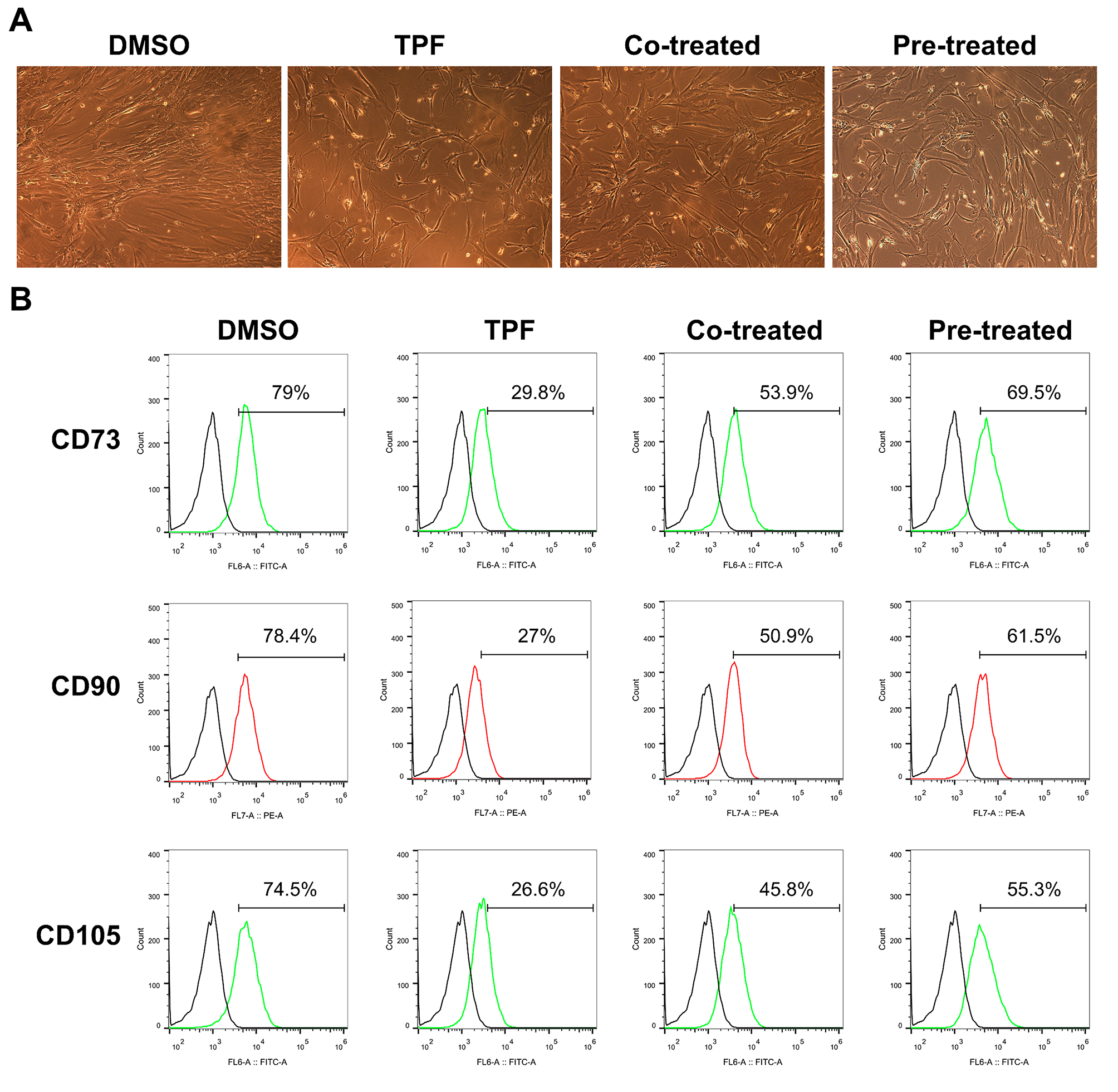
Anti-CCA activity evaluation
To evaluate the anti-CCA activity (tumor inhibition, survival time prolongation, and metastasis prevention) of the CMC-AL on tumor growth, the human CCA cell line CL-6 was used for tumor xenografting in nude mice. The cell was kindly provided by Associate Professor Adisak Wongkajornsilp, Department of Pharmacology, Faculty of Medicine, Siriraj Hospital, Bangkok, Thailand. CL-6 cells were cultured with RPMI 1640 medium and were removed from the culture flask by cell scraper. All cells were collected in a 15 ml conical tube and centrifuged at 100×g for 5 min (25 oC). Cell supernatant was removed and resuspended in 3 ml of complete medium. The cell number was counted using hemocytometer chamber. Cells for injection (1,000,000 cells/200µl complete medium) were prepared and injected subcutaneously into the right upper flanks of nude mice following disinfection of the injection site [19]. Mice were observed daily, and tumor size and body weight were measured every two days before the experiment.
Mice were randomly allocated to three dose groups, i.e., 1,000, 3,000, and 5,000 mg/kg body weight based on the MTD of AL [19]. The control groups were treated with cisplatin and a control vehicle. Six mice per group were allocated to each group and matched-paired according to tumor size (after tumor nodules reached the volume of approximately 50–100 mm3). Animals were fed daily with all test substances by intragastric gavage for 30 days.
Endpoint parameters
Tumor growth inhibition: Tumor growth inhibition was evaluated in all animals. Two linear dimensions were measured as maximum longitudinal diameter (length) and greatest transverse diameter (width) every two days during the investigation period, using a digital external calliper (Mitutoyo, Kawasaki, Japan). Tumor volume was determined using the formula: Tumor volume (mm3) = (length × width2)/2.
Survival time prolongation
The dates of death after treatment were recorded in all mice. Mice were sacrificed with CO2 euthanasia [15] when the growing tumor burden impaired their locomotion, altered vital signs like respiration, caused failure to eat or drink, and other activities [20]. The median survival time (days) of CCA-xenografted nude mice receiving CMC-CCA at all dose levels and reference controls were compared.
Tumor metastasis inhibition
Following euthanasia, autopsies were performed to identify macro-metastases in all animal groups. In addition, primary tumors and organs (lungs, kidneys, heart, liver, brain, spleen, and thymus) were harvested, washed with normal saline and fixed with 10% NBF for histopathology processing (H&E staining) to identify tumor metastasis. The morphological changes within the primary tumor and distant metastases to organs/tissues of the control and treated groups were observed under a binocular compound microscope with the camera (Leica Microsystems, Wetzlar, Germany) at 100x (oil immersion) and lower magnifications.
Immunoblotting
VEGF levels in tumor cells and tissues were detected using a human VEGF ELISA kit (Cat. no. ab100663: Abcam, UK) according to the manufacturer’s instructions. Lung tissues were homogenized using a homogenizer. All reagents, samples and standards were prepared and equilibrated to room temperature (25 °C). Standard or samples were added to each well of the 96-well ELISA plate. Biotin antibody was added to each well, followed by streptavidin solution. TMB One-Step Development Solution was added to each well and incubated at room temperature. Finally, stop solution was added, and absorbance (OD) was measured at 450 nm using a Varioscan™ flash microplate reader machine (Thermoschientific, MA, USA).
Statistical analysis
Statistical analysis was performed using SPSS software version 18.0 (SPSS Inc., Chicago, USA). Qualitative data are presented as numbers (n) and/or percentages (%). Quantitative data are presented as median with 95% confidence interval (CI) values. Differences between two or more quantitative groups of data were performed using Mann-Whitney U-test and the Kruskal Wallis test (followed by pair-wise comparison), respectively. Kaplan-Meier analysis was applied to the survival data of mice in each group. The statistical significant difference was set at α < 0.05 for all tests.


留言 (0)