Mushroom materialsProcurement and authentication of mushroom material
The fruiting body of Leucopaxillus gentianeus was obtained in October 2021 from a rural location in Maharashtra's Sangli District and verified by Dr. M. V. Kale, the head of the botany department at Jaysingpur College in Jaysingpur, Maharashtra, India.
Collection and processing of mushroom material
The harvested mushroom material was naturally dried in the shade and put through an electrical grinder to reduce its size. The resulting powder was sieved before being employed in a further extraction procedure.
Chromatographic and chemical used
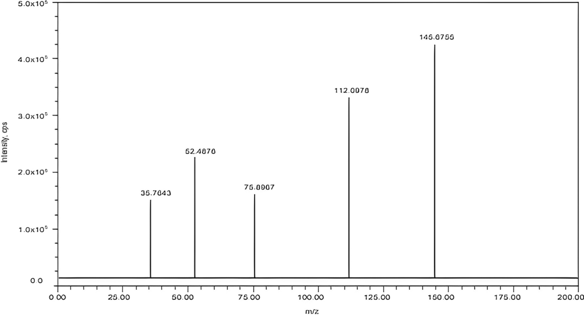
Shimadzu HPLC is comprised of one pump, ultraviolet detector, and LC software to analyze the sample. A mixture of Methanol and water (80:20) was used as a mobile phase. This solution was degassed in a sonicator during the pre-examination phase. The used column for analysis was the C18 column and C8, with the protective column. The flow rate was adjusted at 0.8 ml/min, injecting 10 μl of samples by auto-sampler. All extract samples were detected by a UV detector at 230 nm wavelength. The room temperature was below 25 °C.
Ethanol, Petroleum ether, Chloroform, and Ethyl acetate were obtained from Merck, and progesterone and estrogen were obtained from Sigma Chemical Co. (Sigma-Aldrich). All other chemicals and solvents were in analytical grade.
ExtractionPreparation of mushroom extract
The continuous hot Soxhelation method was used for successive extraction of the fruiting body of Leucopaxillus gentianeus with Til oil, Pet. Ether, Chloroform, Ethyl Acetate, Methanol, and Water, according to their polarity.
Extraction of the fruiting body of Leucopaxillus gentianeus by using Til oil
100 g powder of dried fruiting body of Leucopaxillus gentianeus was packed in a Soxhlet extractor. 1000 ml of til oil was added to the round bottom flask and the maintained temperature was 90–100 °C by using a heating mantle. Extraction was carried out with til oil for several cycles till a drop of solvent from the syphon tube did not leave a greasy spot on the filter paper after evaporation. It took approx. 15 cycles and it requires approx. 4 days. After completion of the process, the marc was taken out from the extractor and was spread as a bed on a clean filter paper and naturally dried till the whole solvent got evaporated. 93 g of dried marc was obtained and used for further extraction with petroleum ether solvent.
Extraction of the fruiting body of Leucopaxillus gentianeus by using Petroleum ether
93 g powder of dried fruiting body of Leucopaxillus gentianeus was packed in a Soxhlet extractor. 1000 ml of petroleum ether was added to the round bottom flask and the maintained temperature was 90–100 °C by using a heating mantle. Extraction was carried out with petroleum ether for several cycles till a drop of solvent from the syphon tube did not leave a greasy spot on the filter paper after evaporation. It took approx. 8 cycles and it requires approx. 35 h. After completion of the process, the marc was taken out from the extractor and was spread as a bed on a clean filter paper and naturally dried till the whole solvent got evaporated. 88 g of dried marc was obtained and used for further extraction with chloroform solvent.
Extraction of the fruiting body of Leucopaxillus gentianeus by using Chloroform
88 g powder of dried fruiting body of Leucopaxillus gentianeus was packed in a Soxhlet extractor. 1000 ml of chloroform was added to the round bottom flask and the maintained temperature was 90–100 °C by using a heating mantle. Extraction was carried out with chloroform for several cycles till a drop of solvent from the syphon tube did not leave a greasy spot on the filter paper after evaporation. It took approx. 10 cycles and it requires approx. 41 h. After completion of the process, the marc was taken out from the extractor and was spread as a bed on a clean filter paper and naturally dried till the whole solvent got evaporated. 83 g of dried marc was obtained and used for further extraction with ethyl acetate solvent.
Extraction of the fruiting body of Leucopaxillus gentianeus by using Ethyl acetate
83 g powder of dried fruiting body of Leucopaxillus gentianeus was packed in Soxhlet extractor. 1000 ml of ethyl acetate was added to the round bottom flask and the maintained temperature was 90–100 °C by using a heating mantle. Extraction was carried out with ethyl acetate for several cycles till a drop of solvent from the syphon tube did not leave a greasy spot on the filter paper after evaporation. It took approx. 10 cycles and it requires approx. 41 h. After completion of the process, the marc was taken out from the extractor and was spread as a bed on a clean filter paper and naturally dried till the whole solvent got evaporated. 79 g of dried marc was obtained and used for further extraction with methanol solvent.
Extraction of the fruiting body of Leucopaxillus gentianeus by using Methanol
79 g powder of dried fruiting body of Leucopaxillus gentianeus was packed in a Soxhlet extractor. 1000 ml of methanol was added to the round bottom flask and the maintained temperature was 90–100 °C by using a heating mantle. Extraction was carried out with methanol for several cycles till a drop of solvent from the syphon tube did not leave a greasy spot on the filter paper after evaporation. It took approx. 13 cycles and it requires approx. 54 h. After completion of the process, the marc was taken out from the extractor and was spread as a bed on a clean filter paper and naturally dried till the whole solvent got evaporated. 74 g of dried marc was obtained and used for further extraction with water solvent.
Extraction of the fruiting body of Leucopaxillus gentianeus by using Water
74 g powder of dried fruiting body of Leucopaxillus gentianeus was packed in Soxhlet extractor. 1000 ml of water was added to the round bottom flask and the maintained temperature was 90–100 °C by using a heating mantle. Extraction was carried out with water for several cycles till a drop of solvent from the syphon tube did not leave a greasy spot on the filter paper after evaporation. It took approx. 12 cycles and it requires approx. 48 h. After completion of the process, the marc was taken out from the extractor and was spread as a bed on a clean filter paper and naturally dried till the whole solvent got evaporated. 66 g of dried marc was obtained.
Distillation of mushroom Extract
After completion of the extraction process solvent was removed by distillation and the concentrated extract obtained was dried naturally.
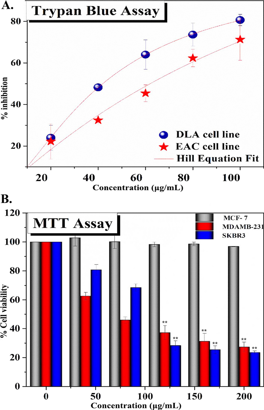
Determination of anti-breast cancer activityHPLC method for mushroom extract
Using HPLC to check extracts purity and efficacy for cancer activity, 1 mg of each extracted sample dissolve in respective solvent extract and run in HPLC.
Pharmacological Activity of mushroom extractProgesterone and estrogen activity with each mushroom extract
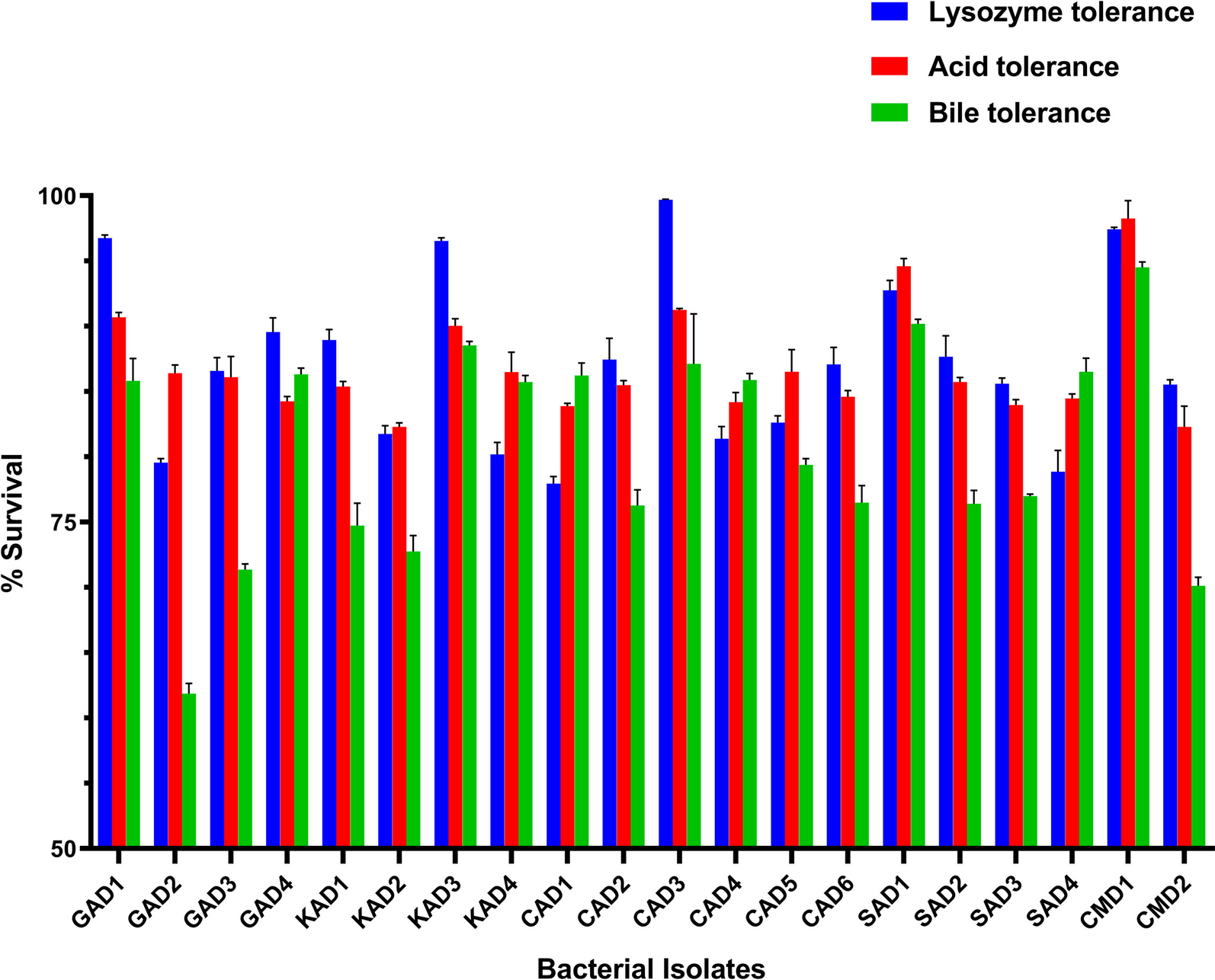
Progesterone and estrogen activity was studied as per different review literature, briefly; 1 mg/ml extract mix with 1 mg/ml concentration of progesterone and estrogen solvent. The reaction was done using the sonication method and absorbance was measured at 254 nm. The control was without any extract used as a positive control. Progesterone and estrogen activity was checked based on the change in peak height.
Screening of cucurbitacin content using TLC and HPTLCTLC
Analyses using thin-layer chromatography (TLC) for both samples Til Oil extract were examined using TLC. Using the dimensional ascending approach, the TLC analysis was performed. Scissors were used to cut a 20 × 20 cm TLC plate covered with silica gel 60G F254 (Merk, India) into a 14 × 3 cm form. After that the plate was lightly chalked with a pencil 1.5 cm away from the bottom and top.
The sample was placed on the TLC plate using glass capillaries at the bottom line that had been pencil-marked. The sample was then loaded once more until a black spot was formed, after which the plate was dried in the fume hood. Then, 20 ml of the solvent (81:11:8) ethyl acetate, methanol, and water was added to the chamber. The plate was inserted into the top of the chamber's liner. Plates were utilized to find the spots after being dried in the fume hood following the run.
Detection of the spot:
After drying each plate, spots were found using UV light at 254 and 289 nm. The retention factor (R f) represented how the active chemical moved.
Rf = Distance traveled by solute/Distance traveled by the solvent
HPTLCPreparation of standard solution
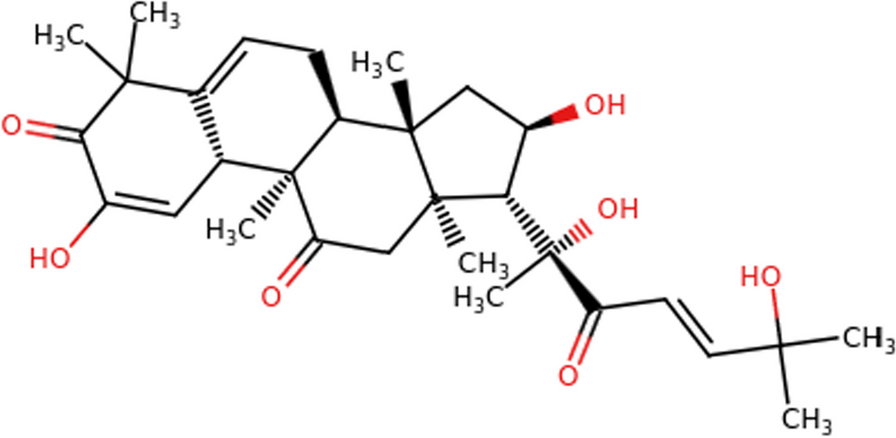
10 mg of precisely weighed cucurbitacin B was dissolved in 10 ml of methanol to create a stock solution of cucurbitacin B (1 mg/ml). A standard solution of cucurbitacin B (40 μg/ml) was created by further diluting the stock solution with methanol. The HPTLC method's working standard was this concentration.
Sample preparation
100 mg of Luffa echinata and Leucopaxillus gentianeus dry fruit and fruiting body powder extracted with til oil was precisely weighed into a volumetric flask of 50 ml. It was sonicated into suspension in 10 ml of methanol. This solution was pipette into a 10 ml volumetric flask in a volume of 2 ml, and 8 ml more methanol was added to make a total volume of 10 ml. This led to the preparation of the sample's stock solution, which had a concentration of 0.4 mg/ml (0.4 g/l). The measurement of cucurbitacin B from the dried fruit powder of the plant material was taken using this concentration. A 0.22 μ membrane filter from Millipore was used to filter all samples.
Instrumentation and chromatographic conditions
In this investigation, the stationary phase was made up of HPTLC aluminum plates that had been pre-coated with silica gel F60 254 and measured 20 × 10 cm with a 200 m thickness (E. Merck, Germany). 254 Before chromatography, the plates were pre-washed with methanol and activated at 110 C for 10 min. Using a Camag Linomat V (Switzerland) sample applicator and a Camag 100 µl syringe, the samples were spotted in the shape of 8 mm bands. The two bands were separated by 12 mm at a constant application rate of 100 nl s°. With a scanning speed of 20 mm/second and a data resolution of 100 m/step, the slit dimension was retained at 6 mm 0.45 mm. The composition of the mobile phase was: ethyl acetate: methanol: and water. A twin-trough glass chamber filled with the mobile phase and used to conduct the linear ascending development. Optimization of chamber saturation. The mobile phase lasted for 30 min at 25 2 °C (room temperature). The chromatogram runs measured 80 mm in length. The plate was then let to air dry at ambient temperature. On the HPTLC plates, the separated bands were scanned from 200 to 400 nm in wavelength. The tungsten lamp was used as the radiation source. At 289 nm, the greatest absorption was discovered. The pictures were taken with the CAMAG Linomat 5 "Linomat5_171103" S/N 171103 (1.00.12) software.
Calibration curve of cucurbitacin B
Cucurbitacin B (0.4 g/l) was produced as a stock solution in methanol. To obtain concentrations of 40, 60, 100, 120, 160, 200, and 240 ng of cucurbitacin per spot, different volumes of standard solution were spotted on the HPTLC plate. Least-square regression was applied to the data of the peak areas plotted against the corresponding concentrations.
Specificity
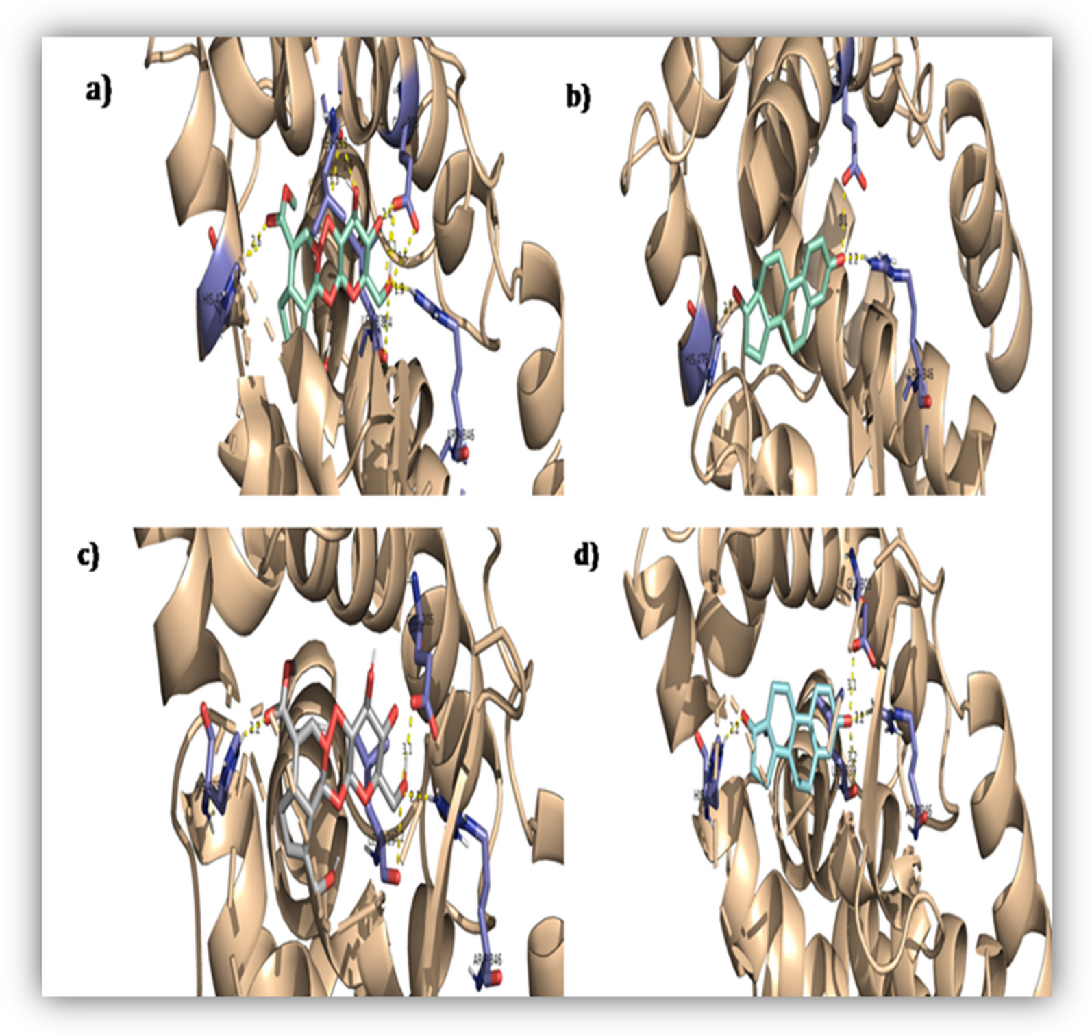
The standard medication and extract were examined to determine the method's specificity. By contrasting the Rf values, the presence of cucurbitacin B in the sample was confirmed. Comparing the spot's measurements and spectrum to the standard. By comparing the spectra at three different levels, namely the peak start (S), peak apex (M), and peak end (E) positions of the spot, the peak purity of the cucurbitacin was determined.
Statistical analysis
All data on anti-progesterone and anti-estrogen activity tests were the average of triplicate analyses. Data were recorded as mean ± standard deviation. Significant differences between means were determined by the Student's test, p values.


留言 (0)