Experimental animals
BALB/c mice and KM mice were used in the experiments. Experiments using specific pathogen free (SPF) grade male BALB/c mice were approved by the Institutional Animal Care and Use Committee of Third Military Medical University (approval number SYXK2017-0002) and were performed in accordance with relevant guidelines. These mice (weighing 18—22 g, 6—8 weeks old) were obtained from Beijing HFK Bioscience Co., LTD (Beijing, China) and housed in a pathogen free environment and fed with free access to food and water. The environment was controlled with the room temperature maintained at 22 ± 2 °C and artificial light–dark cycles of 12 h.
SPF grade KM mice (male, 4—6 weeks old, weighing 18—22 g) were purchased from Hunan SJA Laboratory Animal CO., LTD (Hunan, China). The mice were fed in an individually ventilated cage (IVC) grade animal house of the experimental building of Zunyi Medical University, with free access to feed and water, and were maintained on a 12 h light/dark cycle at 22.0 ± 2.0 °C, with a humidity of 60.0 ± 5.0%. All protocols and experiments procedures involving live animals were approved by the Animal Care Welfare Committee of Zunyi Medical University (approval number SYXK2021-0003).
Cells and culture
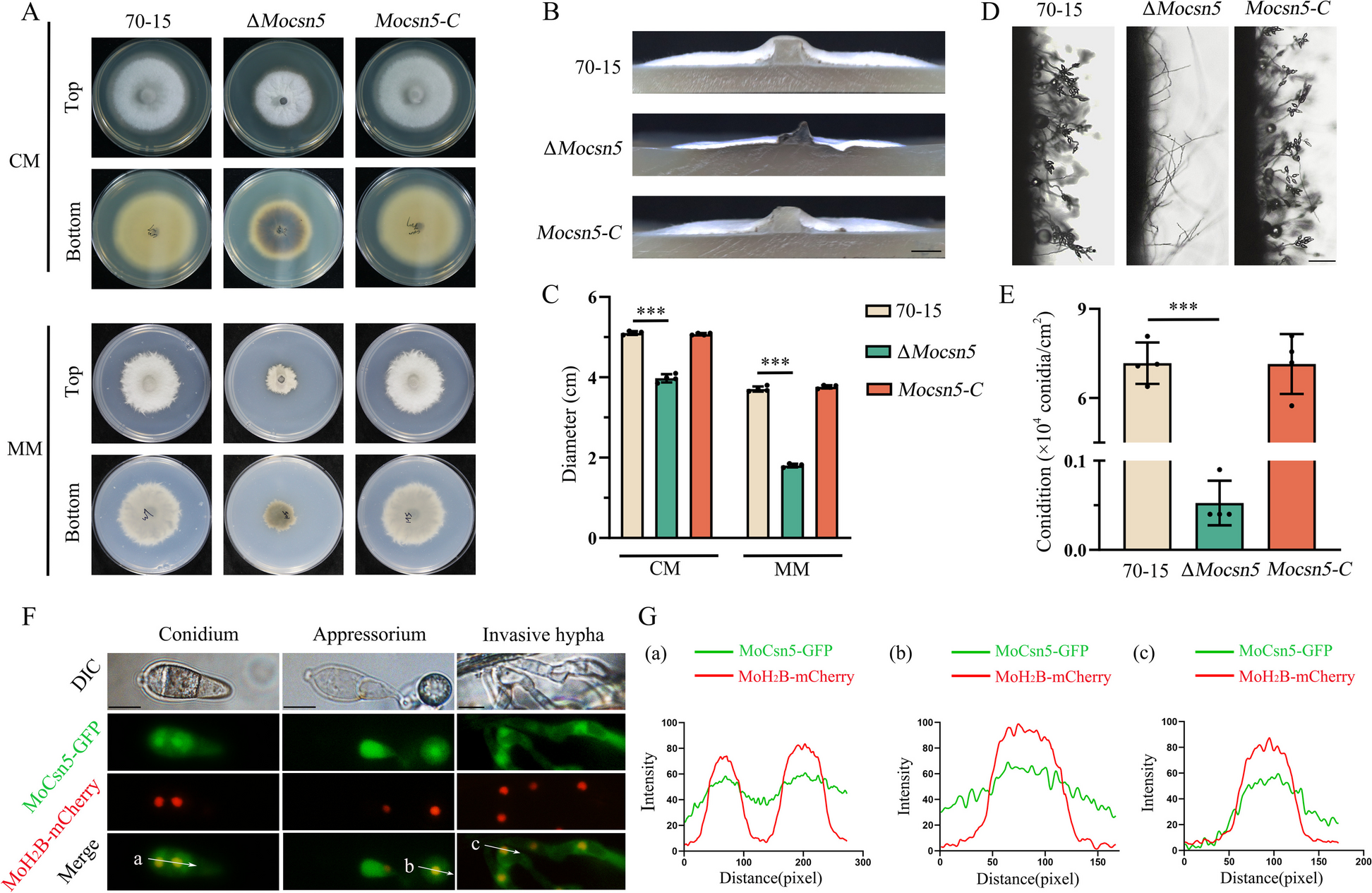
Mouse peritoneal macrophages (PMs) were isolated from male KM mice. The mice were intraperitoneally injected with 3 mL of 3% Thioglycolate (Sigma-Aldrich, St. Louis, MO, USA) on the first day, and the cells were isolated after anesthesia on the third day. The mice were injected with 5 mL of normal saline (NS), gently rubbed for 2—3 min, the peritoneal supernatant was collected, centrifuged, and the supernatant was discarded, and the peritoneal cells were suspended in fresh Dulbecco’s modified Eagle’s medium (DMEM) without fetal bovine serum (FBS) in cell culture dishes at 37 °C in 5% CO2.
Bacterial strain and preparation of bacterial suspension
The PA clinical insolate was kindly provided by Prof. Peiyuan Xia (Southwestern Hospital, Chongqing, China). Bacteria cultured in Mueller–Hinton Broth during the logarithmic phase were collected and diluted in sterile normal saline to achieve a concentration of approximately 1.0 × 10⁸ colony-formation units (CFU)/mL.
Establishment of the LPS-tolerant mouse model and artesunate treatment
To establish the LPS-tolerance model, mice were randomly divided into two groups (10 mice/group) and injected intraperitoneally with LPS (0.3 mg/kg/day) for 3 days, followed by intravenous injection with LPS (50 mg/kg). The survival rate and body weight of the mice were recorded for 7 days. In another experiment, model mice were anesthetized using isoflurane (Keyuan Pharmaceutical, Shandong, China) inhalation after the last LPS injection. Blood, lung and spleen tissues were collected for analysis.
For AS treatment experiments, LPS-tolerant mice were intramuscularly injected with AS (10 mg/kg) at 0 and 4 h after the last LPS injection. At 12 h after the last LPS challenge, blood, lung and spleen tissues were collected for analysis.
Establishment of the second hit (bacterial challenge) mouse model and AS treatment
LPS-tolerant mice or normal mice were intraperitoneally injected with PA (the second hit) at 6 h after the last LPS challenge and the survival rate of the mice was observed for 7 days. For AS treatment, LPS-tolerant mice were intraperitoneally injected with the lethal bacterial dose, and AS was injected intramuscularly at 0, 4, and 24 h after the last LPS injection. The survival rate of the mice was observed for 7 days.
To investigate the effect of AS on the bacterial load, LPS-tolerant mice were treated as described and blood, lung, and spleen tissues were collected at 6 h for CFU count assays.
Establishment of the LPS tolerance cell model
LPS tolerance is used extensively to simulate the sepsis-induced immunosuppression phase in vitro [15]. Herein, a cell model of LPS tolerance was established in mouse PMs. Briefly, cells were cultured with LPS (0111: B4, 5 ng/mL, Sigma-Aldrich) for 4 h. Then, the culture supernatant was replaced followed by the addition of LPS (100 ng/mL) to establish the LPS-tolerance cell model. After an additional 4 h, the culture supernatant was collected, and then TNF-α, as the marker of the formation of the LPS-tolerance model, was assayed.
The influence of a lipid raft inhibitor on the LPS-tolerance cell model
PMs were pretreated with LPS (5 ng/mL) for 4 h, then incubated with LPS (100 ng/mL) and methyl-β-cyclodextrin (MβCD, 5 mM) (MedChemExpress, Shanghai, China) for an additional 4 h. The supernatant was collected to detect the TNF-α level, which is considered to be a marker for the formation of the LPS-tolerance model.
The influence of anti-VDR antibodies on the effect of artesunate
Seven anti-VDR antibodies from different manufacturers (Cell Signaling Technology (12550S); Proteintech (67,192–1-IG); Santa Cruz Biotechnology (SC-13133); Boster (BA2877-2); ABclonal Technology Co., Ltd. Wuhan, China (A2194); Abcam (Ab109234), Cambridge, UK; and Bioworld Technology, Minneapolis, MN, USA (BS91492)) were used to observe alterations is AS’s effect to increase the TNF-α release from LPS-tolerant cells. Briefly, PMs were treated with the seven anti-VDR antibodies (anti-VDR antibody: DMEM = 1:100) for 1 h, separately, and then the PMs was pretreated with LPS (5 ng/mL) for 4 h, incubated with LPS (100 ng/mL), with or without AS (injection preparation, Guilin Pharma Corp, Guangxi, People's Republic of China National Medicine Standard H10930195), and anti-VDR antibodies for an additional 4 h. Finally, the supernatants were collected to detect TNF-α level.
The influence of VDR wild-type and mutant peptides on the effect of artesunate
The two peptide sequences from human VDR (shown in Table 1) containing histidine 397 (wild-type peptide H397) and histidine 305 (wild-type peptide H305) and two mutant peptide sequences with histidine 397 mutated to aspartic acid (mutant peptide H397D) and histidine 305 mutated to alanine (mutant peptide H305A) were synthesized (ChinaPeptides Co., Ltd, Shanghai, China). Briefly, PMs were pretreated with LPS (5 ng/mL) for 4 h, and then AS was added with LPS (100 ng/mL) and the peptides simultaneously. After incubation for another 4 h, the supernatant was collected to detect TNF-α level.
Table 1 Wild and mutant polypeptides sequences from human VDRsiRNA transfection in vitro
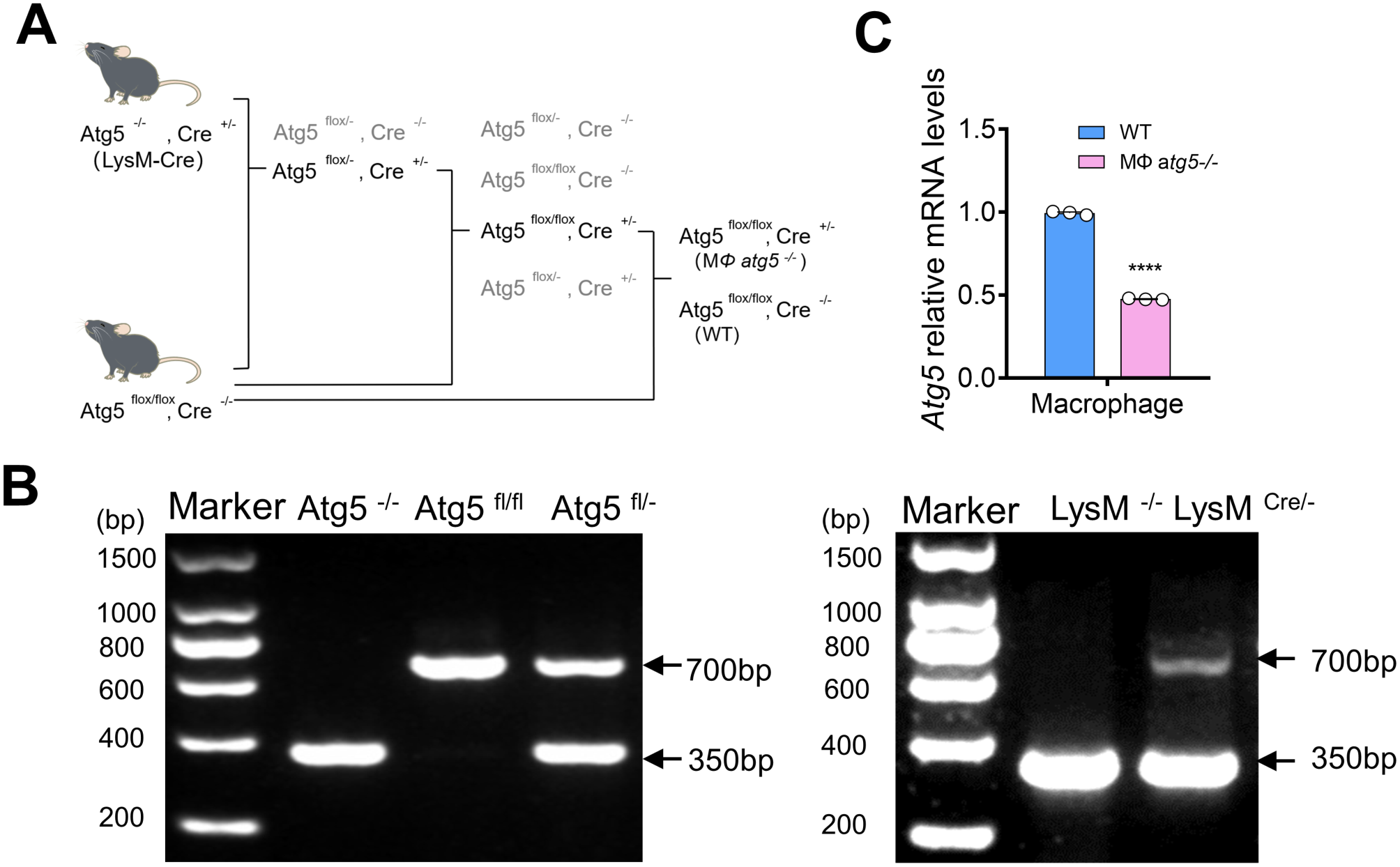
PMs were transfected with an siRNA targeting Cav1 (encoding caveolin-1) or a control siRNA using siRNA Transfection Reagent (SANTA) for 6 h. The medium was discarded and the PMs were incubated for a further 24 h with medium supplemented with 20% FBS and 2% antibiotics. The medium was discarded, and the cells were harvested after incubating again for 24 h with medium containing 10% FBS and 1% antibiotics, then the medium was discarded. The PMs were pretreated with LPS (5 ng/mL) for 4 h, then incubated with LPS (100 ng/mL) for an additional 4 h. The supernatant was collected to detect the TNF-α level, and the PMs were collected to detect the level of ATG16L1 using western blotting.
Molecular docking
The amino acid sequences of human VDR (NP_001017535.1) and mouse VDR (NP_033530.2) were downloaded from GenPept (www.ncbi.nlm.nih.gov/protein), and the crystal structures of the human VDR-1α,25(OH)2D3 complex (1db1) was obtained from the Protein Data Bank (PDB; www.rcsb.org). The homologous 3D structures of mouse VDR were modelled in the ORCHESTRA program of SYBYL-X2.0 by using the known crystal structures of human VDR as the reference. Molecular docking was carried out using SYBYL-X2.0 employing the Surflex-docking program to investigate the detailed interaction between AS and human VDR. The initial binding pocket of AS was subsequently characterized to be close to HIS397 and HIS305 according to a known crystal structure of the human VDR-1α,25(OH)2D3 complex [21]. The interactions of human VDR with 1α,25(OH)2D3 and human VDR with AS, the alignment of different VDRs, and visualizations were performed using an education version of the PyMol package (www.pymol.org).
Binding assay
To confirm the binding of AS to mVDR, cell membrane proteins were extracted, and then mVDR in the extracts was captured to the 96-well plates coated with anti-VDR antibodies (Wuhan Fine Biotech, Wuhan, China). After another anti-VDR antibody (Cell Signaling Technology, USA) targeting another epitope was added to the plate, FITC-labeled AS (Xi'an Rui Xi Biotechnology Co., Ltd., Xi'an, China) was added to the plate and the fluorescence values was finally tested.
Immunofluorescence assay
For cell membrane VDR detection, VDR was labeled with green fluorescent fluorescein FITC. The cell membrane was stained with the red fluorescent DIL, vimentin, or an anti-CD64 antibody.
For cytoskeletal membrane dye labeling, PMs were collected to fixed using 4% paraformaldehyde for 1 h at room temperature, then blocking was performed with goat serum for 1 h at room temperature. The PMs were incubated overnight (over 16 h) at 4 °C with anti-VDR antibody (Proteintech, Wuhan, China), followed by incubation with FITC-goat anti-mouse IgG secondary antibodies (Boster, Shanghai, China). The cells were then incubated with DIL (Solarbio), and anti-vimentin antibody (Beyotime Biotechnology, Shanghai, China) for 10 min at room temperature, respectively. Nuclei were stained using 2-(4-amidinopheny l)-1H-indole-6-carboxamidine (DAPI) (Boster) for 10 min at room temperature. Finally, the cells were photographed under a laser confocal microscope (Leica, Heidelberg, Germany) to observe VDR on the cell membrane.
For red fluorescent anti-CD64 antibody labeled cell membranes, after fluorescent labeling of VDR, PMs were again blocked with serum and incubated with the anti-CD64 antibody (Santa Cruz Biotechnology) or anti-CD64 antibody's control antibody mouse IgG (Beyotime Biotechnology) overnight at 4 °C, followed by incubation with Alexa Fluor 594 (594) goat anti-mouse IgG secondary antibody (Boster). Nuclei were stained using DAPI. Finally, the cells were photographed under a laser confocal microscope (Leica) to observe VDR on the cell membrane.
For cytoplasmic and nuclear VDR detection, PMs were fixed with 4% paraformaldehyde for 1 h at room temperature, permeabilized for 10 min in 0.3% Triton X-100 (Solarbio), resuspended in phosphate buffered saline (PBS). Blocking was performed with goat serum for 1 h at room temperature. The PMs were incubated overnight (over 16 h) at 4 °C with anti-VDR antibodies (Proteintech), followed by incubation with FITC sheep anti-rabbit IgG secondary antibodies (Boster). The nuclei were stained using DAPI (Boster) for 10 min at room temperature. Finally, the cells were photographed under a laser confocal microscope (Leica) for the expression of molecules in the cytosol and nuclei.
Immunohistochemistry
Sections were deparaffinized and hydrated, and then heat-induced epitope retrieval was performed with Citrate Antigen Retrieval Solution (pH 6.0) for 15 min at 100 °C. The sections were peroxide blocked for 10 min, then blocking was performed with goat serum for 1 h at room temperature, then incubated overnight (over 16 h) at 4 °C with anti-VDR antibody (SANTA), followed by incubation with Enhanced enzyme-labeled goat anti-mouse IgG polymer (Zsbio, Beijing, China) for 30 min, Then further treated with the Biotin-Streptavidin HRP Detection Systems (Zsbio) for 15 min, DAB for 10 min, and a hematoxylin counterstain for 5 min. Sections were dehydrated through a series of ascendingethanol concentrations and xylene, neutral gum mounting, finally, photographed under a light microscope (Olympus, Tokyo, Japan).
Extraction of membranal, cytoplasmic and nuclear proteins
Membrane proteins were extracted from PMs using a cell membrane protein extraction kit (Thermo Fisher Scientific, Waltham, MA, USA). Cytoplasmic and nuclear proteins from PMs were extracted using a nucleoprotein Extraction Kit (Solarbio).
Western Blotting
Total cellular protein from PMs was extracted using radioimmunoprecipitation assay (RIPA) buffer (Cell Signaling Technology). The proteins were quantified using a BCA Protein Quantification Kit (GENEray, Shanghai, China). Ten micrograms of each protein sample were separated using sodium dodecyl sulfate polyacrylamide gel electrophoresis (SDS-PAGE) and transferred to a polyvinylidene fluoride (PVDF) membrane. The membranes were blocked with 5% skim milk and incubated with different antibodies [anti-VDR (Santa Cruz Biotechnology, Dallas, TX, USA), anti-ATG16L1 (Cell Signaling Technology)] overnight (over 16 h) at 4 °C. The membrane was washed three times for 10 min each time using Tris-buffered saline containing 0.1% Tween 20 (TBST), incubated with the corresponding secondary antibodies, and washed again. Immunoreactive protein bands were imaged using a chemiluminescent gel imaging system (Bio-Rad, Hercules, CA, USA) and analyzed using Image Lab software (Bio-Rad).
Enzyme-linked immunosorbent assay (ELISA)
Serum, cell culture supernatants and tissue homogenates were collected and the levels of TNF-α, IL-6 and IL-1β were detected using respective ELISA kits (Thermo Fisher Scientific). The VDR protein level was detected using a VDR ELISA Kit (JiangLai Biology, Shanghai, China; Wuhan Fine Biotech, Wuhan, China).
Statistical analysis
All experiments were repeated more than three times, and all values are presented as the mean ± standard deviation (SD). Statistical analysis was performed and plotted using one-way analysis of variance (ANOVA) followed by two tailed unpaired Student's t-test using GraphPad prism 8.0.2 software (GraphPad Inc. La Jolla, CA, USA). In the figures, *, P < 0.05, significant statistical difference; **, P < 0.01, significant statistical difference; #, P > 0.05, no statistical difference.


留言 (0)