2.1 Identification of differentially expressed genes in ccRCC
To reveal the transcriptomic signature of ccRCC, the microarray datasets GSE66270 and GSE68417 from the Gene Expression Omnibus (GEO) database were used [14]. The GSE66270 dataset contains 14 normal and 14 tumor samples. The GSE68417 dataset contains 14 normal and 29 tumor samples. By using the GEO2R tool analysis, genes showing fold change (FC) > 2 and P < 0.05 in their expression in ccRCC as compared to that in normal renal tissues were identified and defined as differentially expressed genes (DEGs) in ccRCC. The overlapping components were analyzed by Funrich software. The ClusterProfiler software package was used for pathway analysis with the Kyoto Encyclopedia of Genes and Genomes (KEGG) database [15]. Next, The Cancer Genome Atlas (TCGA) data portal was used to obtain gene expression profiles of 530 ccRCC patients, and these profiles were analyzed by the limma package [16].
2.2 Cell culture
ccRCC cell lines (A498, caki-1, 769P, 786-O, and OS-RC2) and the immortalized human kidney cell line HK-2 were purchased from American Type Culture Collection. The cells were cultured in RPMI 1640 medium (Biological Industries) containing 10% fetal bovine serum (FBS). For inhibitor treatment, the cells were stimulated with the ATAD2-specific inhibitor BAY-850 (1 µM, MCE) for 24 h. For c-Myc inhibition experiments, the c-Myc inhibitor 10,058–F4 (100 mmol/L, Selleck) was used to treat ccRCC cells for 24 h.
2.3 Synthesis of small interfering RNA, plasmid construction, and cell transfection
The cells were transfected with specific small interfering RNAs (siRNAs) targeting ATAD2 (si-ATAD2). si-ATAD2 was purchased from Biosynthesis (Beijing, China). The sequences of siRNAs were as follows:
si-ATAD2–1 sense: 5′-GAAUAUUGAUAGUAGGAGATT-3′, antisense: 5′-UCUCCUACUAUCAAUAUUCTT-3′;
si-ATAD2–2 sense: 5′-CUAUACCACUAGUGAGAAATT-3′, antisense: 5′-UUUCUCACUAGUGGUAUAGTT-3′;
si-ATAD2–3 sense: 5′-CUGAUAAAGAGGCUCGAAATT-3′, antisense: 5′-UUUCGAGCCUCUUUAUCAGTT-3′;
si-N. C sense: 5′-UUCUCCGAACGUGUCACGUTT-3′, antisense: 5′-ACGUGACACGUUCGGAGAATT-3′.
The ATAD2 gene was cloned into the pcDNA3.1 plasmid. According to the manufacturer’s protocol, lipofectamine 2000 (Invitrogen) was used to transfect the plasmid or siRNA. In the in vivo experiment, the recombinant lentiviral plasmid and the packaging plasmid were co-transfected into 293T cells to generate lentiviral particles. After A498 cells were infected with lentivirus, 4 µg/mL puromycin was used for 48 h for selection. The cells were maintained in a medium containing 2 µg/mL puromycin to obtain a stable cell line.
2.4 Cell proliferation analysis
Cell counting kit 8 (CCK-8) and 5-ethynyl-2′-deoxyuridine (EdU) assays were used to evaluate the changes in cell proliferation. A total of 2 × 10³ ccRCC cells were inoculated in 96-well plates, and CCK-8 (Beyotime) assay was performed after 1, 2, 3, 4, and 5 days. The EdU kit (Ribobio) was used for EdU measurement in accordance with the manufacturer’s instructions.
2.5 Western blotting assay and antibodies
Proteins were separated using 8–10% sodium dodecyl sulfate-polyacrylamide gel electrophoresis (SDS-PAGE). The separated proteins were transferred to polyvinylidene difluoride (PVDF) membranes (Millipore, Billerica, MA, USA). After blocking with a 5% blocking solution, the PVDF membranes were incubated with primary antibodies overnight at 4 °C. Following incubation with the appropriate secondary antibodies, the Luminata Crescendo Western HRP substrate (Millipore) was used for processing, exposure, and digital imaging.
Supplemental Table S1 provides the list of antibodies used in the experiment.
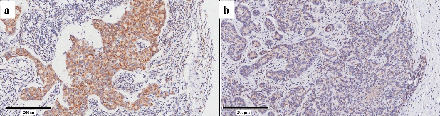
2.6 Immunohistochemistry and immunofluorescence analysis
Antigens were retrieved from deparaffinized tissue sections, which were then blocked with 5% bovine serum albumin. Tissue sections were incubated with primary antibodies overnight at 4 °C, followed by incubation with peroxidase-conjugated secondary antibodies to detect antigen-antibody complexes. Subsequently, a color reaction was performed using a 3,3ʹ-diaminobenzidine (DAB) substrate kit (ZsBio). ccRCC cells were plated on coverslips and fixed in 4% paraformaldehyde (PFA) for immunofluorescence staining. Next, the cells were treated with 0.25% Triton X-100 for 15 min. After blocking with 5% donkey serum, the coverslips were incubated overnight with primary antibodies at 4 °C. Fluorescent secondary antibodies were used to detect the primary antibodies.
2.7 Immunoprecipitation
IP lysis buffer (20 mM Tris-HCl, 150 mM NaCl, and 1% Triton X-100, pH 7.5) was used for cells lysed. The protein lysis buffer was incubated with antibodies (anti-ATAD2, anti-c-Myc, or IgG as control antibodies) overnight on a shaker at 4 °C, followed by incubation with protein A + G agarose beads (Santa Cruz) for 4 h. Immune complexes were eluted from the agarose beads and subsequently analyzed by SDS-PAGE followed by immunoblotting assay. Light chain-specific secondary antibodies (1:2000, 58,802 S; CST) were used to prevent the IgG heavy chain from obscuring the signal of the target protein. The Flag-ATAD2 and HA-c-Myc plasmids were co-transfected into HEK293T cells by Lipofectamine 2000 (Invitrogen). After 48 h, the cells were lysed with IP lysis buffer. Flag fusion proteins were immunoprecipitated by anti-Flag magnetic beads (Beyotime) and eluted with 3X Flag peptide (Beyotime) and analyzed by SDS-PAGE followed by immunoblotting.
2.8 In vivo animal studies
This study was approved by the Ethics Committee of Peking University Third Hospital (A2023031). A subcutaneous xenograft tumor model was used to evaluate the effects of ATAD2 on tumor growth in vivo. A total of 5 × 10⁶ vector control cells and cells stably expressing ATAD2 were injected subcutaneously into the armpits of nude mice. After 1 week, the subcutaneous tumor volume was calculated with a digital caliper every 7 days by using the following formula: V = width2\(\times\) length/2. Mice were monitored, at minimum, once every 3 days, and the tumors were not allowed to exceed 1.5 cm in diameter or 1500 mm3 in volume. After 4 weeks, the mice were euthanized. The tumor tissues were then weighed and paraffin-embedded for hematoxylin and eosin (H & E) staining and immunofluorescence analysis.
2.9 Silver staining
The Flag-ATAD2 plasmid was transfected into HEK293T cells. The IP lysis buffer was used to prepare the whole cell lysate. Subsequently, the whole cell lysate was immunoprecipitated overnight with Flag magnetic beads or IgG control beads (Beyotime) at 4 °C. The precipitate was then washed with cold IP cleaning buffer for 5 times. In accordance with the manufacturer’s instructions, the rapid silver staining kit (Beyotime) was used to visualize the isolated ATAD2 binding proteins.
2.10 Detection of glucose, adenosine triphosphate, and lactic acid levels
Glucose, lactic acid, and intracellular adenosine triphosphate (ATP) levels were identified with the corresponding detection kits (Nanjing Jiancheng Corporation) in accordance with the manufacturer’s instructions.
2.11 c-Myc green fluorescent protein reporter assays
To monitor c-Myc activity, a c-Myc green fluorescent protein (GFP) reporter plasmid was obtained from Yeasen (11744ES03). The sequence of the c-MYC response element was as follows: GGCCTAACTGGCCGGTACCGCTAGCCTCGATCACGTGCACGTGCACGTGCACGTGGCGCGTAGATCTGCAGAAGCTTAGACACTAGAGGGTATATAATGG.
The c-Myc GFP reporter plasmid, and the ATAD2 plasmid were co-transfected into HEK293T cells for 48 h. The c-Myc pathway activation was evaluated by detecting the GFP with a fluorescence microscope.
2.12 Statistical analysis
All experiments were conducted at least three times. The data are expressed as mean ± standard deviation (SD). Data analysis was conducted using GraphPad Prism 8.0. Statistical analysis was performed using unpaired two-tailed t-test, two-way ANOVA, and one-way ANOVA followed by Tukey’s multiple comparison test. A P-value of < 0.05 was considered statistically significant.


留言 (0)