
記住我


The progressive improvement and increased availability of software-based methods for quantitative analysis of radiological images are driving a significant shift in the way radiologists analyze images. The application of such methods on chest CT images is constantly expanding, particularly in the management and risk stratification of lung nodules (both solid and subsolid) [4, 5, 12, 13, 21,22,23,24,25,26,27, 30,31,32,33,34,35,36,37,38,39,40,41].
Currently, software-based analysis of lung nodules is very promising and is attracting great interest, not only among thoracic radiologists but also among other thoracic physicians. Among software-based methods applied to SSNs, those related to the prediction of histological invasiveness of resected SSNs are widely described in the literature [35,36,37,38,39], whereas those related to the prediction of growth are few and have been investigated almost exclusively in Asian populations [22,23,24,25,26,27, 30, 41].
To our knowledge, the present study is the first to evaluate the performance of a software-based method for predicting the growth trend in solitary NSNs with a diameter of 6 − 30 mm in a Caucasian (Italian) population.
In our study, we tested the predictive value of several quantitative CT features such as Feret’s diameter, perimeter, area, mean CT attenuation, median CT attenuation, modal CT attenuation, standard deviation of CT attenuation, LMD, skewness of CT attenuation, kurtosis of CT attenuation, shape descriptors (circularity and solidity). These quantitative CT features were extracted from the largest cross-sectional areas of the NSNs on baseline CT images using an open-source software (ImageJ). From the data obtained using the ImageJ software, we found a significant correlation between NSN growth and the following CT features: Feret’s diameter, perimeter, area, LMD, skewness of CT attenuation, kurtosis of CT attenuation, circularity, and solidity. This significant correlation was found both by considering the absolute DT value and by comparing the groups of growing and nongrowing NSNs based on the DT cutoff value of 1,556 days. The univariate analysis confirmed the value of the same CT features for predicting future NSN growth; however, in the multivariate analysis, only the skewness of CT attenuation and the LMD were independent predictors for NSN growth. Specifically, a positive skewness value greater than 0.90 and a LMD greater than 19.16 mg/mm were significantly associated with NSN growth (Fig. 5).
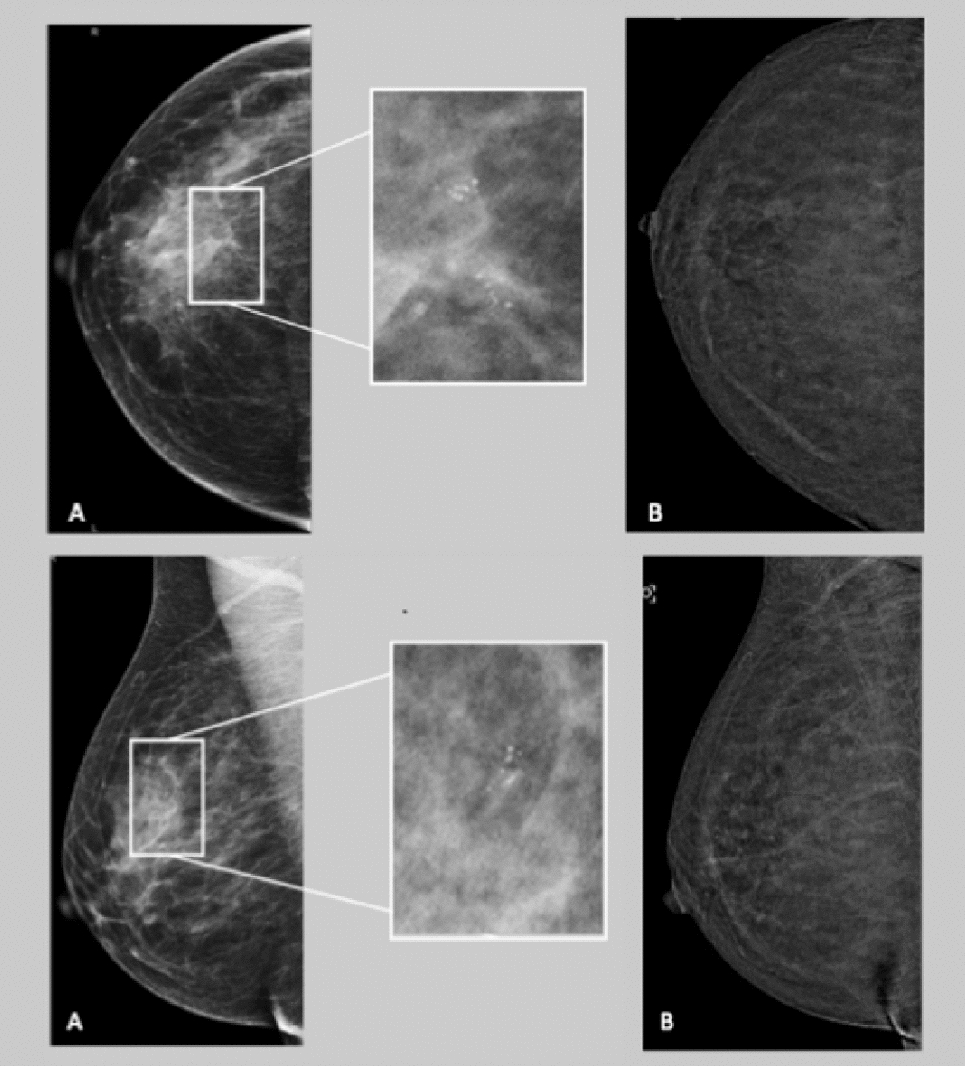
Fig. 5
Growing nonsolid nodule (doubling time of 586 days) in the left upper lobe in a 71-year-old man, former smoker with no previous oncologic history. The interval between the baseline (left) and the last follow-up CT examination (right) was 1020 days. The software-based analysis performed on the baseline CT images obtained a skewness value of 1.61 and a LMD of 29.42 mg/mm. This nodule was surgically excised with a histopathological diagnosis of invasive pulmonary adenocarcinoma graded as pT1cN0
In our study, the skewness of CT attenuation was found to be the strongest predictor of growth, and the two models that included the skewness, with or without LMD, showed an excellent predictive power for NSN growth.
Skewness of CT attenuation is a measure of the asymmetry of the distribution of CT density and represents the distribution pattern of CT attenuation in a histogram. A positive skewness value reflects a histogram with a tail longer on the right than on the left side. The higher positive skewness of CT attenuation observed in growing NSNs is consistent with a greater asymmetry of the CT attenuation distribution. The significant difference in skewness of CT attenuation between growing and nongrowing NSNs found in this study probably reflects the greater intralesional heterogeneity of growing NSNs compared to that of nongrowing NSNs. In other words, even if a pathological correlation was not performed in this study, we assume that the positive skewness observed in the growing NSNs is the result of the presence of some pixels with higher CT attenuation which could reflect the presence of more aggressive neoplastic foci.
The LMD is a quantitative feature that combines density and size measurements in a single value and it represents the two-dimensional variant of the mass [12, 13, 30]. As recently reported [30], LMD is a quantitative feature particularly appropriate to evaluate the characteristics and the behavior of SSNs on CT images, both at the baseline and during follow-up. In a previous retrospective study, Bak et al. [22] found that the 97.5th percentile of the mean CT attenuation and the slope of mean CT attenuation from the 2.5th to the 97.5th percentile, two other histogram-related CT features not tested in our study, were useful predictors of growth in a group of 54 NSNs. More recently, Sun et al. [23] showed that uniformity, another quantitative feature, is an effective predictor of growth in a group of 42 NSNs, with lower uniformity values in growing NSNs. Similar to skewness of CT attenuation, the 97.5th percentile of the mean CT attenuation, the slope of mean CT attenuation from the 2.5th to the 97.5th percentile, and uniformity reflect the intralesional heterogeneity of growing NSNs [22, 23]. Therefore, based on the findings of the present and the above previous studies, it can be stated with reasonable certainty that quantitative CT features reflecting intralesional heterogeneity are useful predictors of NSN growth.
We also found no significant correlation between NSN growth and certain quantitative features related to CT attenuation (i.e., mean, median, modal, and standard deviation of CT attenuation).
In a similar study performed on Asian (Chinese) patients, Shi et al. [24] reported that only the standard deviation of the CT attenuation value (greater than the cutoff value of 50 HU) and the maximum diameter (greater than the cutoff value of 10.2 mm) were independent predictors of growth in a group of 101 NSNs detected in 59 patients. The differences between ours and their findings could be attributable to several factors: the software-based method used (two-dimensional analysis with ImageJ versus volumetric analysis with 3D slicer); the quantitative CT features considered for the analysis; the method for defining the growing group (DT of LMD versus changes in maximum diameter and appearance of solid component); NSN selection criteria (only solitary NSNs were included in our study); patient ethnicity (Italian versus Chinese patients).
Prior studies have also reported that the mean CT attenuation value is an independent predictor of NSN growth [42, 43]; however, more recent studies, including our own, focused on software-based CT analysis of NSNs did not confirm the predictive value of this parameter [22,23,24,25].
Contrarily to previous studies [42, 43], we did not find any significant relationship between NSN growth and certain independent variables, including sex, smoking habits, history of cancer, and NSN location. Although the univariate analysis revealed a relationship between NSN growth, patient age, and the presence of pulmonary emphysema, this association was not confirmed in the multivariable analysis.
This study had some limitations. First, it was performed retrospectively and included a relatively small number of NSNs; however, our inclusion criteria were extremely strict and only solitary NSNs scanned with the same acquisition-reconstruction parameters and the same CT scanner were selected. Second, the software-based analysis was performed by a single radiologist; however, the reliability in measurement of the selected quantitative features was found to be excellent or good. Third, the quantitative CT analysis was performed only on two-dimensional axial images; however, the largest cross-sectional area should be the most representative image for assessing the internal characteristics and shape features of NSNs.
Despite these limitations, our study further highlights the potential role of software-based quantitative CT analysis, specifically the leading role of skewness of CT attenuation (along with the LMD), as a tool to improve risk stratification and devise personalized management plans for patients with pulmonary NSNs.
It is well known that most persistent NSNs exhibit an indolent course and may remain stable for several years [5, 20]; however, some NSNs grow quickly and require surgical removal [13]. Therefore, identifying quantitative CT features capable of early discrimination between growing and nongrowing NSNs is becoming a crucial aspect of radiological analysis to reduce the number of CT examinations and improve the timing of follow-up. From this clinical point of view, the application of software-based CT analysis to evaluate NSNs improve radiologist performance through deeper image analysis, highlighting CT features that cannot be assessed visually.
Obviously, the performance of software-based analysis and the promising predictive power of the skewness of CT attenuation observed in the present study need to be confirmed in future prospective analyses with larger sample sizes. However, we believe that computer-based quantitative CT analyses applied in NSNs (regardless of the software used) will have strong implications in the clinical setting, as they can affect the decision-making process and nodule management.
In conclusion, the results of the present study showed that the skewness of CT attenuation was the strongest predictor of growth in NSNs with an axial diameter of 6 − 30 mm detected in a Caucasian (Italian) population. According to our preliminary data, NSNs with a skewness value greater than 0.90, specifically those with a LMD > 19.16 mg/mm, should require closer follow-up due to their higher growth potential, and consequently, higher risk of becoming a clinically active cancer.
留言 (0)