Study population
A retrospective study was conducted in a tertiary-level teaching hospital (Xuzhou Central Hospital). The study protocol was reviewed and approved by the Ethics Committee of our hospital and conforms to the Declaration of Helsinki.
Between September 2015 and April 2022, twenty-two patients who underwent VA-ECMO support and LUS measurement and with cardiogenic shock were enrolled in the study. Eleven patients had acute myocardial infarction, 8 had fulminant myocarditis, and 3 had heart failure after cardiac surgery. The patients were divided into survivors (n = 16) and nonsurvivors (n = 6).
In our centre, when the following criteria are met, VA-ECMO treatment is required for patients with refractory cardiogenic shock [9]: (1) Persistence or aggravation of tissue hypoxia (extensive skin mottling, anuria, neurological impairment, elevated blood lactate, etc.) despite adequate fluid loading; or (2) sustained hypotension (systolic blood pressure < 90mmHg or mean arterial pressure < 65 mmHg) despite infusion of very-high-dose catecholamines (epinephrine ≥ 0.3 µg/kg/min, dopamine ≥ 15 µg/kg/min, norepinephrine ≥ 0.3 µg/kg/min). The exclusion criteria included the following: (1) ECMO duration less than 3 days; (2) lack of an appropriate acoustic window for LUS determination; (3) complications with pneumothorax; (4) complications with congenital heart disease; or (5) complications with chronic lung disease.
Data Collection
Demographic and clinical data were collected from the hospital records and our organization’s proprietary database. The demographic data included age, sex, and body mass index (BMI). The clinical data included the Acute Physiology and Chronic Health Evaluation II (APACHE II) score, comorbidities, ICU mortality, length of ICU stay, duration of ECMO and duration of ventilation.
Extracorporeal membrane oxygenation
The ECMO setup involved femoral vein catheterization, femoral arterial catheterization and superficial femoral artery catheterization [10]. The femoral arterial cannula (16-18 F for adults, Medtronic) was inserted into the common femoral artery and positioned in the distal aorta. A vein cannula (20-24 F for adults, Medtronic) was inserted into the femoral vein and positioned in the inferior vena cava close to the right atrium. A 7 F catheter was inserted into the superficial artery to prevent leg ischaemia [11]. The blood flow of VA-ECMO was controlled at 80-100ml/kg. The activated clotting time was maintained at 160–220s. An intraaortic balloon pump (IABP) was applied for all patients to maintain haemodynamic stability.
The adult-related indications for ECMO weaning were used as a reference in our centre, which included the following [12]: stable haemodynamic conditions ≥ 2–4 h; dosages of vasoactive drugs such as dopamine and dobutamine < 10 µg/kg/min; oxygen saturation of internal jugular vein > 70%; pulse pressure close to normal; LV ejection fraction (LVEF) > 40%; central venous pressure ≤ 12 mmHg.
Mechanical ventilation
A protective mechanical ventilation strategy was implemented during ECMO for all the patients, and standardized nursing work was performed under the guidance of doctors. The protective mechanical ventilation strategy included [13,14,15] selection of the pressure control (PC) mode and limiting the tidal volume to < 4 ml/kg, the respiratory rate to 6 ~ 20 times/min, the inspiratory peak pressure to 20 ~ 25 cmH2O, the positive end expiratory pressure (PEEP) between 10 ~ 15 cmH2O, and the oxygen intake concentration between 30 − 50%, and the setting of respiratory rate refers to the changes of tidal volume and ECMO airflow to match. The pH value and arterial blood carbon dioxide partial pressure measured by arterial blood gas analysis were maintained within the normal range.
Lung ultrasound score
Based on the scheme proposed by Bouhemad et al [16], the patients’ chests were divided into 12 regions. Each hemithorax is systematically divided into six regions, two anterior, two lateral, and two posterior, according to the anatomical landmarks set by the anterior and posterior axillary lines. Each region is divided into half, superior, and inferior. To perform a comprehensive examination, all adjacent intercostal spaces were explored in each region of interest by sliding the probe along the space. For each explored region, the most severe finding was reported in simple checkboxes according to the following rating: normal: 0; well-separated B-lines: 1; coalescent B-lines: 2; and consolidation: 3. The cumulative lung ultrasound (LUS) score corresponds to the sum of each examined region score (minimum score, normal lungs: 0; maximum score, both consolidated lungs: 36). ANT = anterior; INF = inferior; LAT = lateral; POST = posterior; SUP = superior. All procedures were performed by trained sonographers. For a full inspection with a 13 − 6 MHz linear probe (M-Turbo portable colour ultrasound, Sono), all the adjacent intercostal spaces of each region were explored.
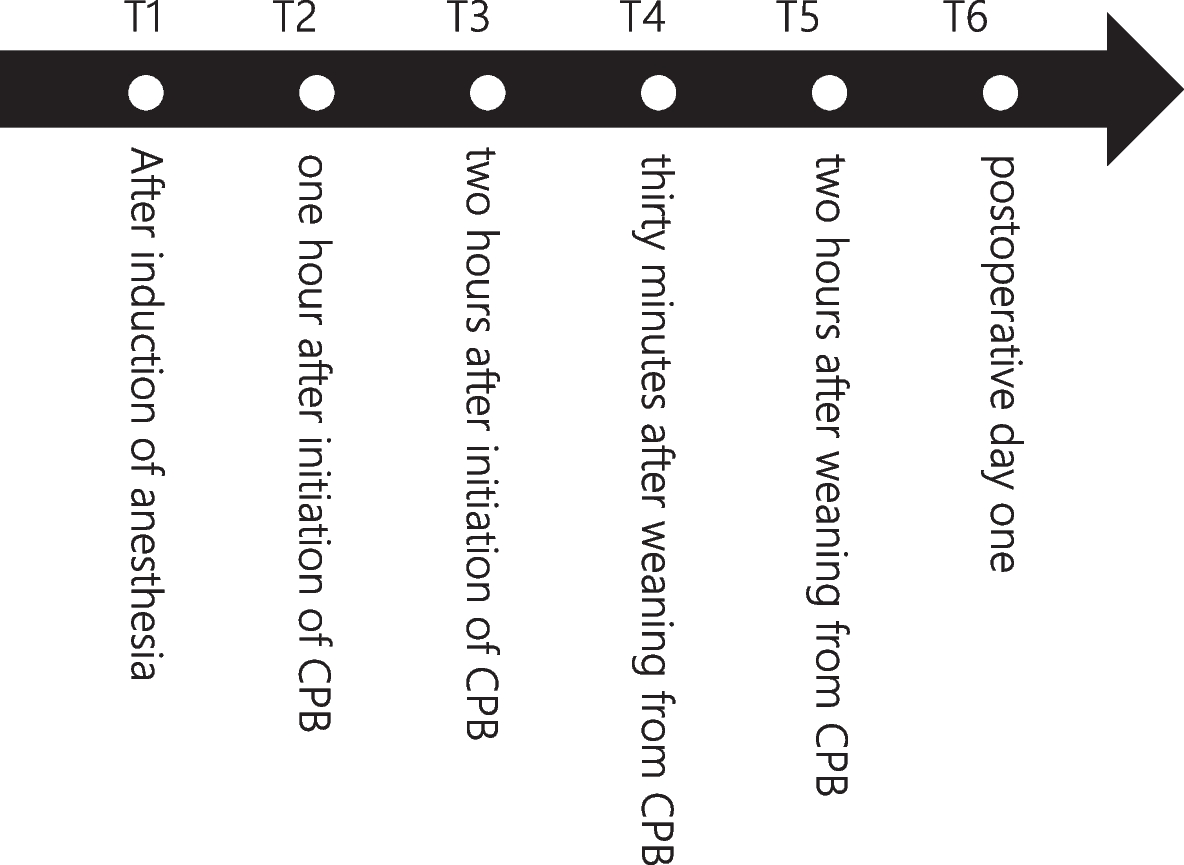
The lung ultrasound scores of the patients were obtained at different periods, including at the initiation of ECMO, each morning in the following four days and at the termination of ECMO and ventilation as T0, T24, T48, T72, T96, TW and TR. According to the above periods, the corresponding ventilator parameters and the blood gas analysis results of the right upper limb artery blood sample were also recorded, including PaO2/FiO2, PaCO2, and pulmonary dynamic compliance (Cdyn).
Statistical analysis
Statistical analysis was performed with IBM SPSS Statistics 26 (IBM Corp., Armonk, NY). Normal distribution was formally tested with the Shapiro–Wilk test. Continuous data are presented as the mean with standard deviation. ANOVA was used to compare repeated measurement data at different time points, and a T test was used for comparisons between two groups. Categorical data are presented as frequencies and percentages. Categorical variables were compared using the chi-square test. The Pearson method was used to compare the correlation between the LUS score and PaO2/FiO2. ROC curves analysis was used to determine the diagnostic value of the significantly changed variables during ECMO support for the ICU survival status of the patients. A p value < 0.05 was considered statistically significant.


留言 (0)