Fluid management is a critical aspect of patient care; however, monitoring technologies for assessing fluid status or the need for administration of fluids have traditionally been restricted to the care settings in the OR and ICUs. The need for effective fluid management tools is significant in other settings. One example is the ER, where invasive technologies such as arterial and Swan-Ganz catheters are rarely found, yet the need for assessing and adjusting fluid status is often critical. A number of non-invasive devices have attempted to fill this void, but introduction has been limited due to device size, technical complexities, patient comfort, cost issues, and accuracy [1, 2]. There remains a need to measure cardiac output (CO) noninvasively by a clinically validated technique; ideally, by means of wireless and small-footprint technologies that can be utilized quickly and effectively in many environments.
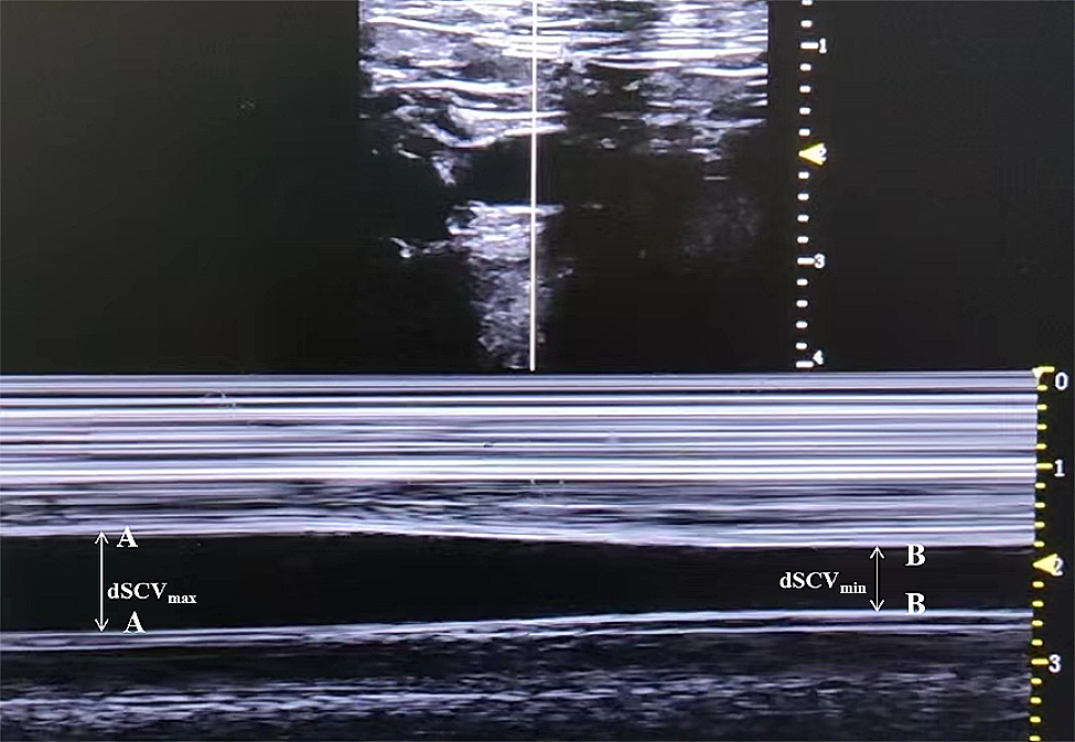
The Vitalstream (VS) continuous noninvasive physiological monitor (Caretaker Medical LLC, Charlottesville, Virginia, further referred to as CTM) is FDA-cleared for the measurement of heart rate, continuous noninvasive BP, and respiratory rate as well as advanced hemodynamic parameters of cardiac output/stroke volume, left ventricular ejection time and heart rate variability. The system and the underlying approach have been described in detail elsewhere [3, 4]. Briefly, the VS tracks central aortic BP via pulse analysis, specifically Pulse Decomposition Analysis (PDA), of the peripheral pulse at a distal site, typically the finger. The device uses a low pressure [30–40 mmHg], pump-inflated, finger cuff that pneumatically couples arterial pulsations via a pressure line to a pressure sensor for detection and analysis. Physiological data are communicated wirelessly to a tablet-based user interface via Bluetooth or Wi-Fi. The device is currently cleared only for adult use, i.e., patients 18 years and older. There are no regulatory restrictions on the clinical use of the device, however, just like all finger-based sensing technologies, extremely cold fingers can impede operation.
The PDA approach is based on the concept that primarily two central reflection sites are responsible for the shape of the pressure pulse envelope of the upper body [5,6,7]. The two reflection sites, one located at the aortic juncture of thoracic and abdominal aortas, and the other at the iliac bifurcation, reflect the primary left ventricular ejection pulse to give rise to two additional, reflected, component pulses that trail the primary ejection pulse, as a result of which, within the pulse pressure envelope of each cardiac cycle, these three component pulses arrive sequentially in the arterial periphery. The model of the spatio-temporal behavior of these three component pulses constitutes the PDA formalism that can be used to monitor hemodynamic states and changes. The PDA model is based on physical assumptions that are readily testable and which coherently explain the structure of the pulse. The purpose of this study was to validate cardiac output readings, provided by the Vitalstream, both in magnitude and trend direction, against Gold Standard measurements obtained using thermodilution (TD) as well as to assess relative time response characteristics.
CTM’s approach to calculating cardiac output (CO) utilizes a linear model that incorporates arterial stiffness estimation, impedance correction, and integration over the “systolic” area of the pressure pulse [8]. A problem with traditional pulse contour approaches has been the estimation of the systolic area, since these approaches frequently simply utilize the “dichrotic” notch to separate systolic and diastolic phases. However, this categorization yields integration over both the actual systolic component pulse area as well as sections of reflected component pulses that complete the pulse envelope, yielding inaccurate systolic area estimates. PDA, which offers a comprehensive and physical explanation of the arterial pulse envelope morphology, provides an opportunity to refine the systolic area calculation.
Setting realistic a-priori expectations, particular in the context of evaluating a non-invasive technology is important as the measurement of cardiac output is a somewhat approximate science. Not only do invasive approaches routinely have performance errors well in excess of 40%, but recent comparisons between Gold Standards have demonstrated similar discrepancies [9]. We refer here to the Fick/TD comparison results reported in Fares, who reported a standard deviation of 2.03 l/min, corresponding to an estimated error larger than 65% [10]. Large discrepancies were also reported by Opotowsky and Tehrani [11, 12]. With these considerations in mind acceptable agreements were errors less than 40% and concordance higher than 0.8.
Such errors can correspond to LOAs on the order of 2.5 l/min, which is highly significant even at an average CO of 5 to 6 l/min. However, this has been the effective state of clinical practice, just one example being the frequently required additional TD boluses to lower the variance of a shot sequence to an acceptable limit. For clinicians needing to make treatment decisions, the potential added benefits with an equivalently accurate but continuous and non-invasive technology like the Vitalstream is the enhanced ability to identify trends and to assess the variability.
In what follows we present the results of comparing the VS cardiac output absolute and trending measurements with those obtained from discrete bolus thermodilution (TD) as well as from continuous cardiac output monitoring (CCO) in patients undergoing cardiac surgery.
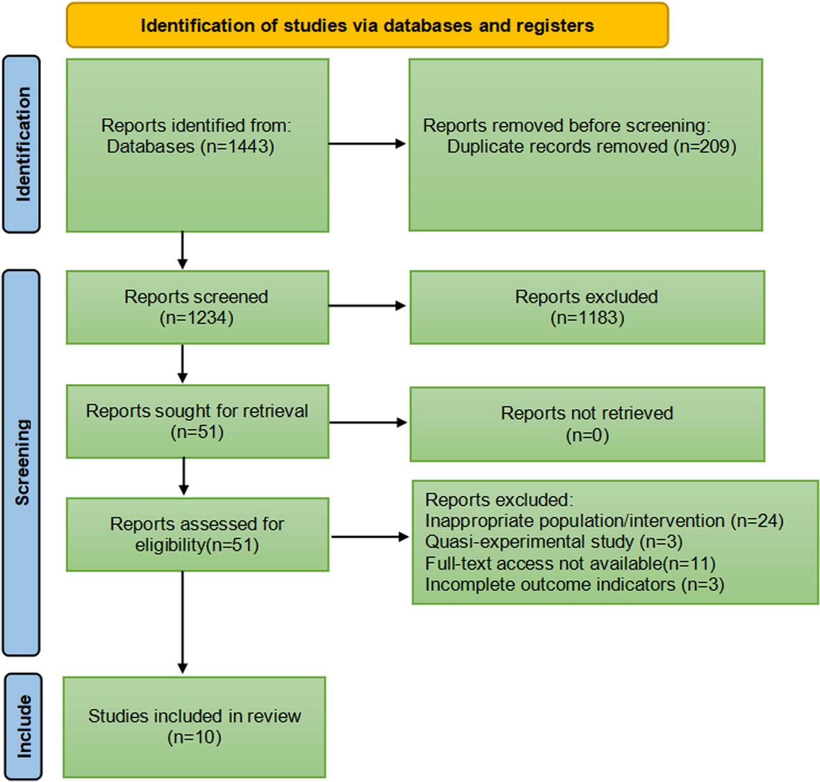
Methods
At Cooper University Hospital patients older than 18y undergoing cardiac surgery in whom pulmonary artery catheter placement was planned as part of their care, were recruited from April 2021 to January 2022 in this IRB approved study. All patients provided written informed consent.
Continuous pulmonary artery catheters (Model #777F8, Edwards Lifesciences Cor., Irvine, CA) were inserted after anesthesia induction. Bolus TD applications were administered depending on clinical indication. For each TD measurement typically three injections were applied within 3–4 min using iced saline via a cold injectate system (Model #93,600, Edwards Lifesciences Cor., Irvine, CA) and averaged. If differences between CO measures exceeded 1 l/min, additional injections were applied. In addition, intermittent semi-real-time cardiac output measurements were obtained using the continuous pulmonary catheter-based cardiac output monitoring system (CCO). The CCO utilizes a heating element placed in the right ventricle that is triggered in a random series of heating bursts, and a thermistor placed in the pulmonary artery for detection of the heat bursts. Since the cross-correlation algorithm that establishes detection requires lengthy input sequences, the system reports changes in cardiac output with delays on the order of 5 min, as reported by others [13]. All TD were performed on the HemoSphere® advanced monitoring platform (Edwards Lifesciences Cor., Irvine, CA). In contrast, the VS’s CO response is significantly faster and primarily driven by the averaging over 30 heartbeats.
The arterial pressure pulse signal was continuously measured noninvasively using the VS device. In order not to interfere with the surgical procedures the device was placed on the patient’s wrist during the procedure setup, with the finger cuff coupled to the middle phalanx of the middle finger, and data transmission was verified, after which time no further physical interaction with the device was possible.
Operation of the device would commence after an initial blood pressure self-calibration procedure, lasting approximately 25 s, during which time the device scans the finger cuff’s coupling pressure from 0 to 250 mmHg while collecting the pressure-modulated arterial pressure pulse signal. At the end of the pressure scan, systolic and diastolic blood pressures are calculated from the processed signal envelope. Thereafter, the device was programmed to perform self-calibration scans at 5-minute intervals, operating in between in the continuous tracking mode with the finger cuff pressure collecting pulse data at a fixed baseline cuff pressure of between 20 and 45 mmHg. The coupling pressure for continuous operation is determined as part of the self-calibration procedure and held constant until the next procedure. Collected data were sent via Wi- Fi interface to an Android tablet for storage.
Data inclusion
With regard to the discrete TD and the CCO data, all data deemed acceptable by the attending clinicians were used in the analysis. In the case of the VS data, a custom signal/noise factor (SNF) was used to identify poor quality data sections which were excluded. The factor is based on the standard ratio of the variances of the physiological signal band to the noise band and obtained using Fourier spectral analysis over an 8-s window with 1 s overlap. The frequency range of the band associated with the physiological signal was set to 1– 10 Hz, based on data by the authors and results by others while the noise band was set to the 100–250 Hz frequency range, which is subject to ambient noise but contains no signal relevant to the base band phenomena of the arterial pressure pulse or its propagation characteristics [14]. Data sections with an SNF below 80 were excluded from the analysis.
Statistical analysis
All comparisons between VS and TD/CCO data were post-processed. In order to match the VS CO readings to the averaged discrete TD bolus data, the averaged CO readings of the ten seconds of VS CO data points prior to a sequence of TD bolus shots was matched. Time alignment was based on the medical record time and the VS time-stamped data points. Likewise, for VS/CCO comparisons ten seconds of VS CO data were averaged bracketing the time stamp of a CCO reading.
A statistical analysis of the data compared the accuracy of the VS absolute CO values with and without initial calibration to the discrete TD CO values, as well as the trending ability, i.e., ΔCO values of the VS physiological monitor compared to those of the reference, specifically using the four-quadrant analysis described by Saugel [15]. The analysis was performed using the MATLAB software package (Natick, USA) and Stata 17.1 (StataCorp, College Station, TX).
The accuracy against reference TD measurements was assessed via correlation analysis as well as via Bland–Altman and difference count distribution analysis of the CO values and standard concordance analysis of the ΔCO values (with a 15% exclusion zone). The Bland–Altman analysis took repeated measurements per subject into account [16]. Separate correlation and Bland-Altman analyses, for calibrated and uncalibrated cases, were also performed on the high and low CO ranges, specifically below 5 l/min and above 8 l/min. A post-hoc Bland-Altman power analysis using the resulting means and standard deviations showed power to be 0.82 for all patients and 0.78 for the 39 patients with complete data.
CCO time delay analysis
In order to access the VS delay in measuring CO with acute changes, an iterative concordance analysis with the “real-time” data provided by the previously described CCO system used at Cooper University Hospital was performed. We follow here the example provided by Saugel, who discusses a method for assessing delays between two methodologies that measure cardiac output [15]. The approach is based on introducing delays between both methodologies’ data and observing concordance results for improvements in trending. The approach of this analysis was facilitated by calculating the cross-correlation spectrum to determine the optimum delay between both cardiac output data series.

留言 (0)