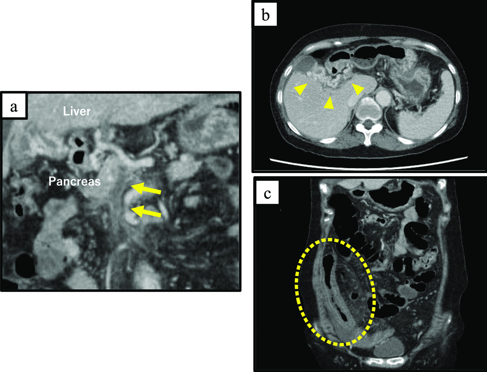
Anatomical variation of the IMA is relatively rare. Kakihara et al. reported that, among 3,182 cases of abdominal angiography, one case (0.03%) had a defective IMA, two cases (0.06%) had abnormal running of the root associated with visceral retroversion, and two cases (0.06%) branched from the SMA. All other cases bifurcated to the left side at the level of the third lumbar vertebra [3]. Several primitive mesenteric arteries arise from the dorsal aorta in a segmental fashion and are distributed against their respective primaries in the early fetal period. Early in development, there is a longitudinal anastomosis connecting the arteries vertically in the dorsal mesentery. According to fetal development, some primitive mesenteric arteries disappear, while the others become the celiac artery, the SMA, and the IMA, which are distributed in the foregut, midgut, and hindgut, respectively [4]. The IMA is initially formed at the twelfth thoracic vertebra but eventually migrates to the level of the third lumbar vertebra [5]. In this case, the IMA bifurcation from the SMA may occur due to both abnormal arterial disappearance and lack of the usual migration. According to the previous reports, the IMA arising from the SMA typically diverges as the first branch supplying the colon (as shown in Table 1) [2, 3, 6,7,8,9,10,11,12,13,14,15,16]. In most cases, the left colic artery arises as a branch of the IMA. One-third of the previous cases were associated with other vascular malformations. In the present case, the IMA was the first branch of the SMA and had the same bifurcation pattern as most previous cases, which included the left colic artery and the sigmoid colic artery. This case also showed a branch from the lumber splanchnic nerve supplying the left-sided colon at the level of the third lumber vertebra where the IMA normally branches. It is possible that the sympathetic nervous system and the vascular system develop asynchronously.
Table 1 Summary of previous cases of the IMA arising from the SMAFor left-sided colorectal cancer with unusual IMA bifurcation, the optimal definition of the extent of lymph node dissection has not been established [3, 13,14,15,16]. Kakihara et al. performed radical lymph node dissection including around the root of the IMA branching from the SMA for cT3N0M0 cStage II lower rectal cancer [3], while Kondo et al. performed central lymph node dissection up to the level of the inferior edge of the duodenum where the IMA usually branches from the aorta, preserving blood flow to the LCA for rectal cancer cT3N0M0 cStage II [14]. Minamisawa et al. dissected the lymph nodes around the bifurcation of the LCA as the cranial border of the dissection for cT2N0M0 cStage I rectal cancer [15]. In this case, preoperative PET-CT suggested metastases in the para-intestinal lymph nodes, but not in the proximal lymph nodes along the IMA. Pathological examination revealed complete resection; therefore, central lymph node dissection including that at the level of the inferior border of the duodenum would be oncologically sufficient. When the IMA was deficient, the marginal artery from the middle colic artery was the only blood flow to the left-sided colon. Thus, regional lymph node dissection in the direction of the intestinal axis is more important than central lymph node dissection in such cases.
Although anatomical variation of the IMA is infrequent, preoperative assessment using CT and CT angiography is very important because they can provide clear case-specific anatomical information. Even in patients with rare anatomical anomalies, prior confirmation of the anatomy allows us to safely complete radical surgery.


留言 (0)