Venous Invasion in Pancreatic Neuroendocrine Tumors Is Independently Associated With Disease-free Survival and Overall Survival
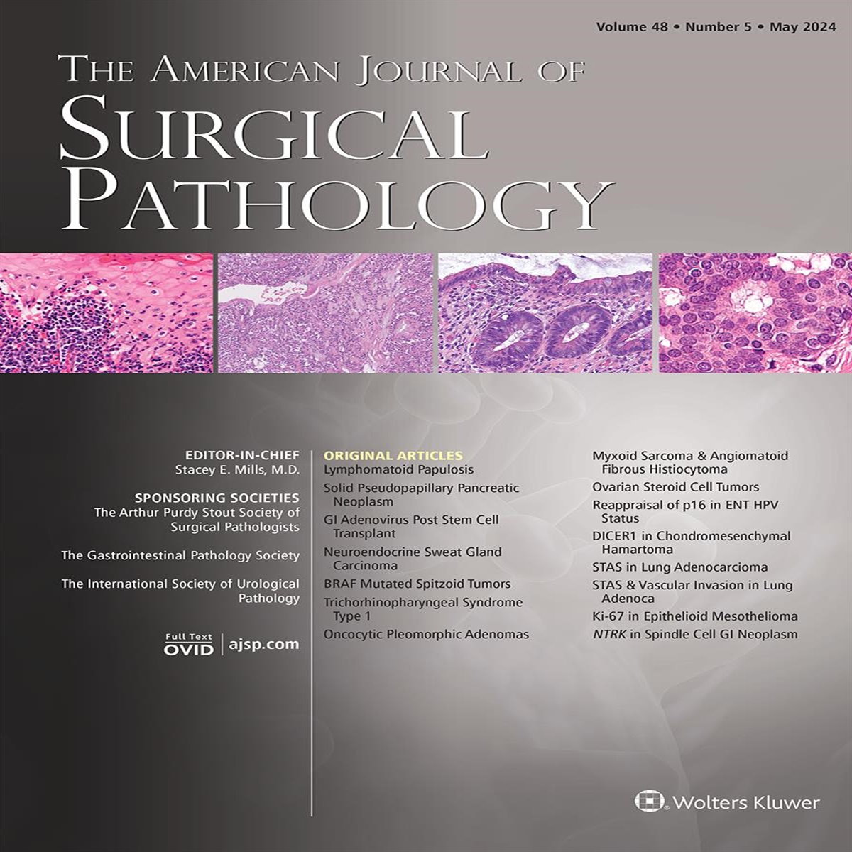
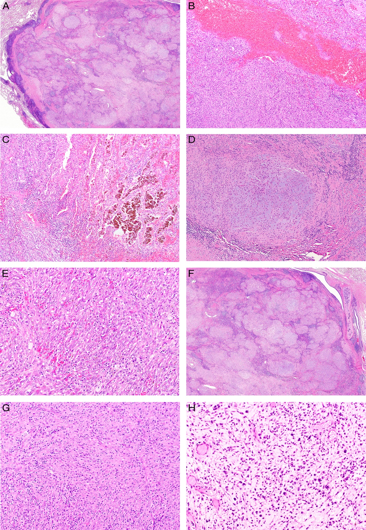
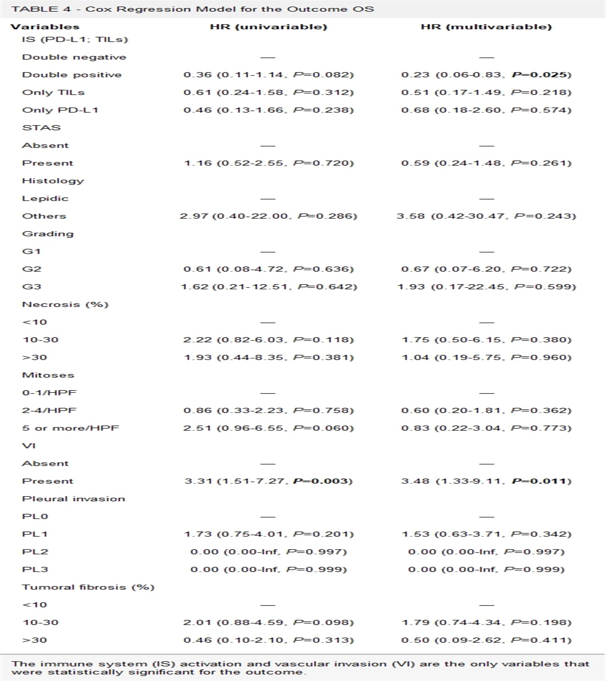
In this study, we evaluated venous invasion and its association with survival in patients with resected pancreatic neuroendocrine tumor (PanNET). Surgical Pathology Archives were searched for pancreatectomies performed for PanNET between October 1, 2005, and December 31, 2019. Hematoxylin and eosin (H&E)-stained slides were evaluated for venous invasion, and Movat’s stain was performed in all cases with no venous invasion detected on H&E stains. Pathology reports and electronic medical records were also reviewed. Venous invasion was identified in 23 of 145 (15.9%) cases on H&E stains, and Movat’s stain identified additional 34 cases with venous invasion (39.3% overall). Orphan arteries with adjacent well-defined tumor nodules or subtle hyalinizing nodules in hyalinizing tumors are highly specific for venous invasion. In stage I-III cases (n=122), venous invasion was associated with larger tumor size, higher World Health Organization (WHO) tumor grade, perineural invasion, extrapancreatic extension, lymph node metastasis, and liver metastasis (P<0.05). In univariate analyses, tumor size, WHO grade, venous invasion, perineural invasion, T stage, and lymph node metastasis all correlated with disease-free survival; however, only venous invasion was associated with worse disease-free survival in multivariate analyses (P<0.01). In all-stage cases, venous invasion was the only attributor associated with worse overall survival in multivariate analyses (P=0.03). In summary, venous invasion in PanNET can be histologically subtle, and Movat’s stain can greatly increase the detection rate. More importantly, enhanced venous invasion by Movat’s stain correlates independently with disease-free survival in patients with stage I-III tumors and overall survival in all-stage patients.

留言 (0)