Plant material and extraction
Jasminum humile L. in the flowering stage was collected on 20th May 2019 from EL-Keram farms, Moderayat al-Tahrir, El-Behaira government, Egypt. It was taxonomically identified by Dr. Mohammed El Gebaly (Consultant botanist-Orman Garden) and a voucher specimen (3.10.16.1) was kept in the herbarium of Faculty of Pharmacy, Cairo University. The plant flowers were collected, washed, and shadow dried. The flowers were then pulverized to a coarse powder (2–3 mm) with an electric blender before extraction. Pulverized flower (50 g) was extracted in a percolator with 80% methanol for 24 h at room temperature. Three washes with 100 mL of fresh solvent with a hold time of 5 h were carried out until the last extract was colorless. The combined extract was filtered Whatman No.1 filter paper and was further concentrated under vacuum using a rotary evaporator at 45 °C to yield 10.5 g dry extract. The crude solid extract was labeled and stored under cold conditions before analysis.
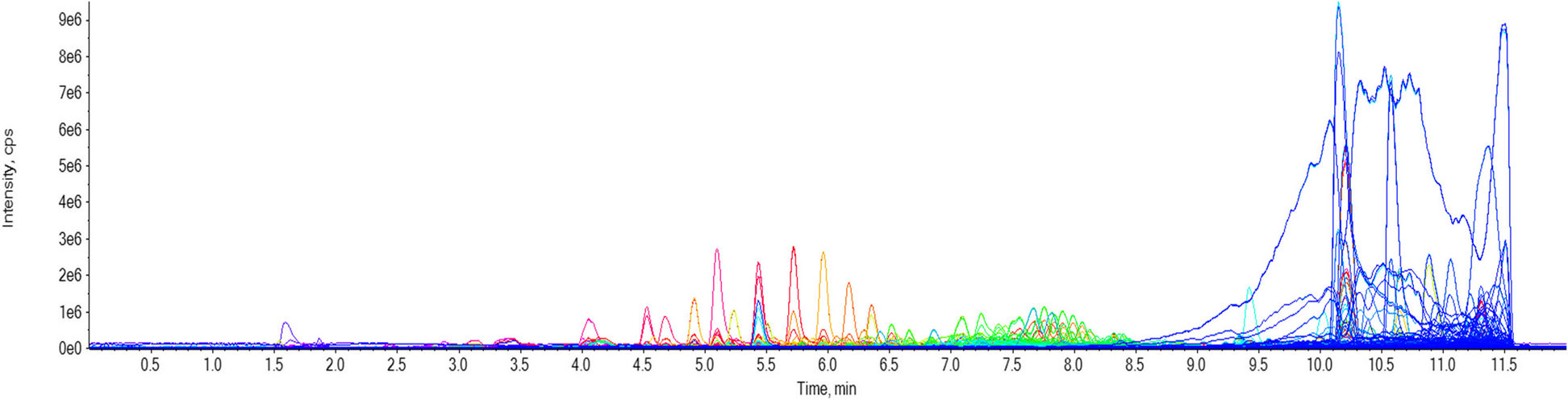
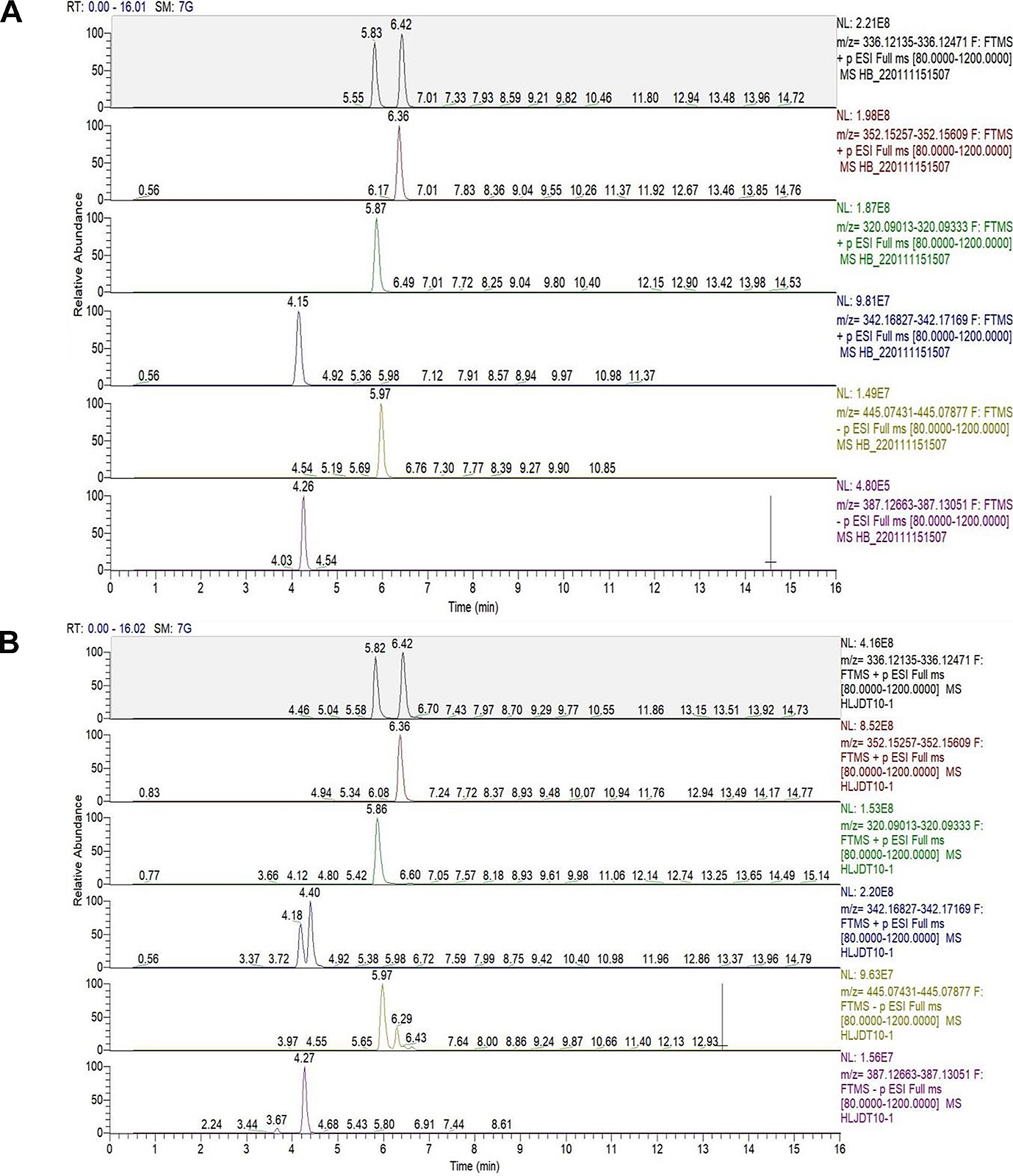
HPLC–PDA-MS/MS
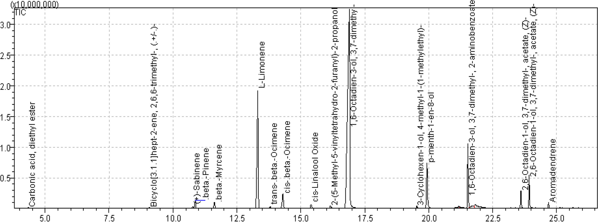
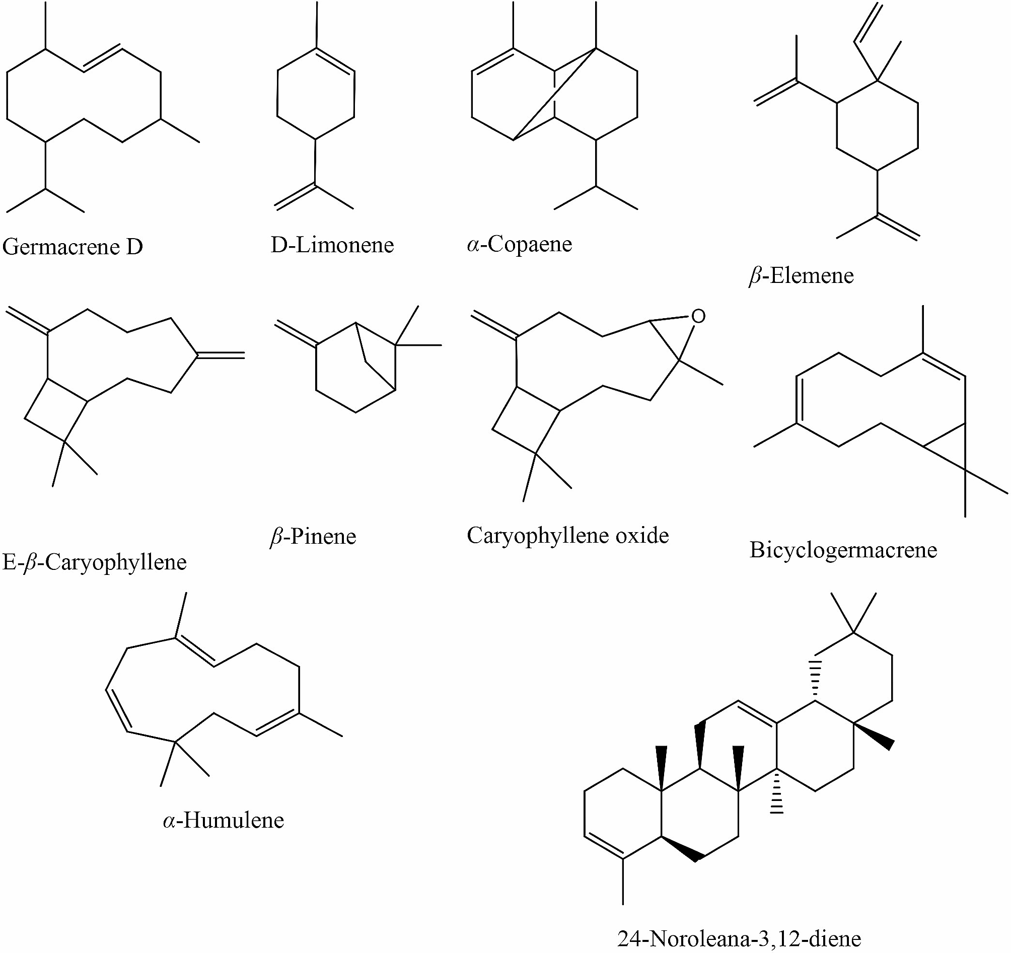
A Thermo Finnigan LC system was used for the HPLC–PDA-MS/MS analysis of J. humile extract (Thermo Electron Corporation, Austin, TX, USA). The instrument utilized was a Zorbax Eclipse XDB-C18, Rapid resolution, 4.6 150 mm, with a 3.5 µm column (Agilent, Santa Clara, CA, USA). Acetonitrile concentration was raised from 5 to 30% in 60 min at a flow rate of 1 mL/min and a 1:1 split before the ESI source using gradient elution with water and acetonitrile (ACN), each containing 0.1% formic acid. Thermo Quest ESI source-equipped LCQ-Duo ion trap was utilized for MS analysis. The system was managed by Xcalibur software (XcaliburTM 2.0.7, Thermo Scientific, Waltham, MA, USA). The MS operating settings were employed in the negative mode [18].
Biological evaluationMTT assay
MCF-7 cells were maintained in RPMI-1640 medium supplemented with 10% FBS, glutamine (2 raM), penicillin (100 units/mL), and streptomycin (100 µg/mL). The cells were cultured at 37 °C in a humidified 5% CO2 incubator. The extract of J. humile flower was tested for in vitro cytotoxicity, using MCF-7 cells by 3-(4,5-dimethylthiazol-2-yl)-2,5-diphenyltetrazolium bromide (MTT) assay. One hundred µL of (RMPI 1640) media was loaded into each of the 96-well plates (triplicate). The final volume for each well was 100 µL. The cultured MCF-7 cells were pooled in a 50 mL vial. Then, the cells were plated at a density of 1 × 10⁶ cells/mL cells/well (100 µL) into 96-well microtiter plates. Each sample was replicated 3 times and the cells were incubated at 37 °C in a humidified 5% CO2 incubator for 24 h. After the incubation period, MTT (20 µL of 5 mg/mL) was added to each well and the cells were incubated for another 2–4 h until purple precipitates were clearly visible under a microscope. Flowingly, the medium together with MTT (190 µL) were aspirated off the wells, DMSO (100 µL) was added, and shake the plates for 5 min. Measure the absorbance spectrophotometry at 540 nm in a microtiter plate reader and the percentage cell viability was calculated manually using the formula:
$$\%\;\mathrm\;\mathrm\:=\:(\mathrm\;\mathrm\;\mathrm\;\mathrm\;\mathrm\;/\mathrm\;\mathrm\;\mathrm\;\mathrm)\:\times\:100$$
A dose–response curve was plotted to enable the calculation of the concentrations that kill 50% of the MCF-7 cells (IC50) compared to the standard drug, etoposide, and the effect of the extract on normal keratinocyte cells (HaCaT).
DNA-flow cytometry analysis
The influence of J. humile extract (5, 10, and 20 µg/mL) on the cell cycle distribution of MCF-7 cell line was evaluated using the CycleTEST™ PLUS DNA Reagent Kit (Becton Dickinson Immunocytometry Systems, San Jose, CA) according to the manufacture instructions. PBMC cells served as control cells with known DNA content for determining the DNA Index (DI) of the inspected samples. Propodium iodide (PI) was used as a DNA-binding dye before running on the DNA cytometer [19]. CELLQUEST software was used for analyzing Cell-cycle distribution.
Annexin V-FITC apoptosis assay
Annexin V-FITC/DAPI assay (Cayman Chemical, Ann Arbor, MI) was used to analyze apoptotic cells. After culturing MCF-7 cells into a monolayer, they were treated with J. humile extract at 5, 10, and 20 µg/mL. Cells were subsequently collected through trypsinization, double washed in phosphate buffer saline (PBS) followed by the binding buffer. Afterward, cells were re-suspended in 100 µL of binding buffer with the addition of 5 µL of FITC-Annexin V (Becton Dickinson BD PharmingenTM, Heidelberg, Germany) followed by a 30 min. incubation period at 4 °C. Cells were then washed in binding buffer and re-suspended in 150 µL of binding buffer with the addition of 1 µL of DAPI (1 µg/µL in PBS) (Invitrogen, Life Technologies, Darmstadt, Germany). The flow cytometer BD FACS Canto II (BD Biosciences, San Jose, CA) was used to analyze the cells and the results were deduced with FlowJo7.6.4 software (Tree Star, FlowJo LLC, Ashland, OR) [20].
Oxidative stress parameters
Spectrophotometric assessment of Superoxide dismutase (SOD) activity at 560 nm following the method of [21] was done based on inhibition of nitro blue tetrazolium- NADH and phenazinemethosulphate (PMS)-mediated formazan formation. Spectrophotometric assessment of Catalase (CAT) was done using the method of [21]. The assay measures the decomposition of H2O2 by CAT at 240 nm. Glutathione reductase (GSH-R) was assessed spectrophotometrically [21] based on the reduction of oxidized glutathione by GSH-R with the help of NADPH. Afterward, the thiol group of reduced glutathione reacts with the chromogen to yield a colored complex measured at 405 nm.
Network PharmacologyTarget Genes Associated with Breast cancer and Selected Compounds
Binding DB (https://www.bindingdb.org/bind/index.jsp) was used using the "homo sapiens" setting to predict target genes for selected compounds based on SMILES. The "minimum needed interaction score" was set to "high confidence (0.700)" during Binding DB prediction. The public database DisGeNET (http://www.disgenet.org/) was used to identify disease-related target genes [22].
Interactions between Compounds and Overlapping Genes: Network Construction
Cytoscape ver. 3.9.1 (https://cytoscape.org/) was used to construct, display, and analyze the network of interactions based on the Binding DB prediction results for constituents and overlapping genes. Nodes in the network indicate bioactive components and genes, while edges show interactions between compounds and genes. Anti-breast cancer components and hub genes were identified by analyzing the network's topological structure and setting the "Degree value" of compounds or genes. A compound's or a gene's degree value represents how many phytoconstituents or genes are present in a network. The therapeutic effects of compounds are enhanced if the compounds target more disease-inducing genes [22].
Building a Protein–Protein Interaction Network
An online database called STRING (https://string-db.org/) was used to gather information on protein–protein interactions between the target proteins of selected J.humile components (PPI). The website calculated a score for each protein's mutual information. The stronger the contact between the two target proteins, the higher the score. Since high-confidence data > 0.7 were used to ensure accuracy and reliability, the study was considered reliable. The obtained protein interaction data was imported into the Cytoscape 3.9.1 application to generate a PPI protein interaction network. The CytoHubba plug-in was employed for the identification of Hub genes [23]. The cyto hubba plg-in has 12 topological features among which we used the “Degree” parameters to screen out the top-ranked proteins according to their degree value of interaction.
Target Protein Gene Ontology and KEGG Enrichment Analysis
It was found that proteins that interact with the active components of J.humile play a role in gene function and signaling pathways by using the database for annotation, visualization, and integrated discovery (David) v 6.8 to evaluate gene function and KEGG pathway enrichment, respectively [24]. The functional enrichment database DAVID, which is accessible online, aids researchers in understanding the bioactivity of a large number of genes. In the current study, p-value ≤ 0.01 was chosen, and the top 10 KEGG pathways and GO enrichments were picked for further investigation. Cellular components (CC), molecular functions, biological processes, and pathways were all included in the list of target proteins. KEGG pathway enrichment results were used to decipher the possible molecular mechanisms of J.humile against breast cancer. KEGG pathway bubble charts made with SRPLOT (http://bioinformatics.com.cn/) were of particular interest.
Molecular docking
The current study used the MOE-Dock from Chemical Computing Group Inc. to carry out computational experiments. The crystal structures of EGFR were obtained using the Protein Data Bank (PDB id 4HJ0). After editing the crystal structures to remove the water molecules and add hydrogen atoms to the protein, MOPAC 7.0 was used to minimize the energy. Then, the alpha spheres produced were used to create dummy atoms by identifying the active site. In docked regions, RMSD values of less than 1.3 were clustered together. The docked complex in the lowest energy-minimized stance was chosen for further investigation. Ten different conformations were carefully chosen. The MMFF94x force field energy computation was then used for the resulting docked complex model to determine the energy parameters and estimate the docked interactions at the active site [25].
Statistical analysis
All data are displayed as the mean ± S.D of three separate determinations. The arithmetic mean and the standard deviation (SD) of all assessments were calculated by the software program Microsoft Excel 2007. Statistical analysis was performed using GraphPad Prism version 5.01. To establish the statistical significance in relation to the reference standard, a one-way ANOVA was employed, followed by a Tukey-posthoc test.

留言 (0)