Collection of plant material
Viscum orientale was collected from the University of Mysore campus (12.3081° N Latitude, 76.6390° E Longitude), Mysuru, Karnataka. The tree was authenticated by Prof. Shivalingaiah, Department of Botany, Maharani’s Science College for Women, Mysore, Karnataka, and the herbarium was deposited in Department of Biotechnology and Bioinformatics, JSS AHER, Mysore, Karnataka, India with voucher number VO15. The leaves of the plant were collected, washed and shade dried for one week. A minimum of one week of shade drying was necessary as the leaves were collected in the month of December-January. Dried material was powdered thoroughly for further extraction.
Chemicals
The reagents employed in the experiment were all analytical grades. DPPH (sigma chemicals) Silver Nitrate (MERCK, Germany), all the chemicals and reagents were procured from padmashri chemicals. Mysuru.
Preparation of plant extracts
Dried samples were extracted continuously using range of organic solvents from non-polarity to polarity (Hexane > Chloroform > Ethylacetate > acetone > Water) and the samples were named as VOHE, VOCE, VOEA, VOAE and VOWE. The extraction was performed by overnight stirring at room temperature and was repeated in each solvent till the sample becomes colorless. Extracted solvents were dried using rotary evaporator and lyophilized and stored at 40C, till further use.
Phytochemical analysis
Plant extracts were assessed for the existence of different phytochemicals namely, flavonoids, alkaloids, saponins, phenols, carbohydrates, glycosides, phytosterols, proteins and terpinoids using standard method [8].
Synthesis of silver nanoparticles
Synthesis of silver nanoparticles at varying stirring time intervals [10]. In a 250ml beaker, 1mM of 100ml Silver nitrate solution was prepared. 10-15ml of leaf extract was added dropwise to the silver nitrate solution with vigorous shaking. The bio-reduced silver nanoparticles solution was run on a UV-Visible spectrophotometer at regular time intervals (0, 1, 2, 3, 4 hours) to evaluate the influence of stirring time on silver nanoparticles formation. Then the graph is plotted by λmax on x-axis and absorbance on y-axis. The synthesized AgNPs were lyophilized, weighed and stored for further use.
Characterization of synthesized silver nanoparticle
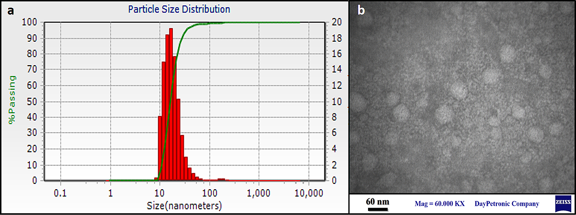
The reduced form of silver on synthesis of nanoparticles were monitored by UV–visible spectra with the range of 200–800 nm, using a UV–Vis spectrophotometer Shimadzu-UV-1800 (Tokyo, Japan) with distilled water as a reference. In the wavelength range 4000–400 cm−1, FTIR spectra were recorded using a Perkin Elmer FTIR RX1 (Dresden, Germany). XPERT-PRO (Bristol, UK)used monochromatic Cu radiation (k = 1.5406 A °) at 40 kV and 30 mA in a 2 h angle pattern for the X-ray diffraction (XRD) examination. Scanning was done between 208 and 808. The crystalline structure was compared to the photographs. FESEM outfitted with an EDAX attachment was used to perform EDAX analysis of silver nanoparticles on a SUPRA55 (CARL ZEISS, Germany). The morphology, size, and shape of the silver nanoparticles were determined via SEM examination. HITACHI H-800 (Tokyo, Japan) SEM tests were performed at 200 kV. A drop of the bio-reduced diluted solution was placed on a carbon-coated copper grid and dried under a lamp to create the SEM grid. Malvern instruments were used to measure the size distribution and stability of AgNPs, as well as DLS and zeta potential [11].
Antioxidant activityDiphenyl-2-picrylhydrazyl radical scavenging activity
Blois (1958) method was used to assess the AgNPs 1,1-diphenyl-2-picryl-hydrazyl (DPPH) free radical scavenging capacity [12]. In several test tubes, different concentrations (20, 40, 60, 80, and 100 μg/ml) of AgNPs and standard butylated hydroxytoluene (BHT) were used. To the above samples, 1 ml of freshly prepared DPPH (0.1 mM) was added and vortexed thoroughly and incubated in dark 30 min. At 517 nm, the absorbance of stable DPPH was measured. As a control, the DPPH without sample was preserved. The inhibition % was used to measure the free radical scavenging activity. The percentage of inhibition was computed, and BHT was used as a reference standard. The percentage inhibition vs. concentration was plotted, and the IC50 value was calculated as the concentration required to inhibit radicals by 50%.
Reducing power capacity
2.5 ml of sodium phosphate buffer was added to the various concentrations of the AgNPs (20, 40, 60, 80, and 100 μg/ml), followed by 2.5 ml of % potassium ferricyanide solution. The content was vortexed well before being incubated for 20 min at 500 °C. Following the incubation period, 2.5 ml of 10% TCA was added to each tube and centrifuged at 3000 g for 10 min. To 5 ml of the supernatant, 5 ml of deionized water was added followed by addition of 1 ml of 1% Ferric chloride and incubated at 350C for 10 min. Absorbance was read at 700 nm. The reference standard was butylated hydroxyl toluene (BHT). The percentage inhibition vs. concentration was plotted, and the IC50 value (concentration required for 50% radical inhibition) was calculated [13].
Nitric oxide radical scavenging assay
Garrat et al. (1964) developed a nitric oxide radical scavenging test [14]. Briefly, 1 ml of 10 mM Sodium nitroprusside was added to all the tubes containing varied quantities of AgNPs (20, 40, 60, 80, and 100 μg/ml), followed by 1 ml of Griess reagent. The reaction mixture was vortexed well and incubated for 1 h at room temperature. In dispersed light, a pink-colored chromophore form. At 540 nm, the absorbance of the mixture was compared to that of the matching blank solutions. The reference standard was butylated hydroxyl toluene (BHT).
Antimicrobial activityPreparation of the bacterial sub-culture
The different bacterial culture used for present investigation namely Escherichia coli, Bacillus cereus, Bacillus subtilis, Staphylococcus aureus and Salmonella typhi and were obtained from P.G. Department of Microbiology, Manasagangotri Campus, University of Mysore, Mysuru. To obtain a bacterial subculture, 100 μl of the bacterial culture was combined with 5 ml of sterile nutrient broth and incubated at 37 °C for 16 h [15].
Antibacterial activity
Using the agar disc diffusion assay method, the antimicrobial activity of the produced silver nanoparticles was assessed. Briefly, autoclaved nutrient agar plates were prepared and to that test bacterial cultures of 100 μl were then swabbed on the surface. Disc diffusion assay was carried out using standard protocol. An antibiotic, Gentamicin is taken as positive control. 10 μg/ml of Gentamicin, 10 μg/ml, 15 μg/ml and 20 μg/ml of the synthesized silver nanoparticles was applied on separate sterile discs of diameter 6 mm (whatman filter paper discs) and allowed to dry before being placed on the agar medium. The plates were incubated at 370C for 24 h and the resulting zone of inhibition was measured [15].
Anthelmintic assay
The anthelmintic assay was performed using the Ajaiyeoba et al., (2001) method [16]. Earthworms (6–9 cm) from the Pheretima posthuma species were collected for this study due to their relatedness and similarities with the human intestinal roundworms. Earthworms were collected from the soil of moist and wet regions of Mysuru farmlands. (https://bmccomplementmedtherapies.biomedcentral.com/articles/10.1186/s12906-016-1219-5).
The collected earthworms were washed with distilled water to remove the fecal matter [17]. Three groups were made with 3 earthworms in each. The time it took to become paralyzed and die was measured. When there is no visible movement of the worms except when they are shaking hard, it is said to be paralyzed. The worms' death time, on the other hand, was recorded when it was confirmed that they did not move when shaken vigorously or dipped in warm water (50 °C).
Hemagglutination assay
In Brief, using microtiter plate, different concentrations of synthesized silver nanoparticles (100 μl) was mixed with 100 μl of 2% suspension of human erythrocyte in phosphate buffer (pH 7.2). The plates were left undisturbed for 1 h for agglutination to take place at room temperature. After incubation time, the results were observed visually [18, 19].
Statistical analysis
Statistical significance was calculated between the groups using two way ANOVA with Turkey’s multiple comparison test. The data was given as a mean ± standard error of the mean (n = 3). Graphpad prism 8.0 was used for statistical analysis.


留言 (0)