Karyomegalic interstitial nephritis, a type of chronic interstitial nephritis, is described when there are enlarged renal tubular epithelial nuclei, interstitial fibrosis, and tubular atrophy. The exact cause of KIN is unknown; however, viral infections, toxic agents, and genetic causes are proposed. Toxic causes include exposure to alkylating agents, heavy metals, and mycotoxins, especially ochratoxin A [7,8,9,10]. KIN has been thought to be hereditary, as about half of cases with KIN had a family history of nephropathy [8]. A familial clustering of disease and increased frequency of human leukocyte antigen (HLA)-A9 and HLA-B35 with genetic defects on chromosome 6 were reported [2, 11]. In addition, KIN has been linked to mutations of the Fanconi anemia associated nuclease 1 (FAN1) gene, which is responsible for DNA repair processes [11,12,13]. Unlike other FAN gene mutations, the FAN1 gene mutation is not associated with Fanconi anemia, risk of malignancy, or developmental anomalies, as the gene is predominant in renal, hepatic, and neuronal tissues [12]. Nevertheless, recent case presentations of a brother and sister with FAN1 mutations and progressive renal failure who developed malignancy after renal transplantation suggest an increased risk of malignancy among patients with kidney transplant recipients and KIN [14]. Recent studies have clarified that KIN is linked to polyploidization of tubular cells, which represents an important mechanism of response to acute kidney injury [15,16,17]. Airik et al. used a mouse model with knocked-out deoxyribonucleic acid (DNA) repair protein FAN1 and showed that FAN1 inactivation resulted in replication stress and persistent DNA damage. Chronic DNA damage causes cells to fail to complete mitosis and undergo polyploidization, which is characteristic of KIN [16]. Considering KIN is a potential hereditary disease, these genetic abnormalities might increase the patient’s susceptibility to viral and toxic factors that contribute to the disease.
Karyomegalic interstitial nephritis is a slowly progressive chronic interstitial nephritis. Clinically, patients present with mild to moderate renal impairment, reaching ESKD in the fourth or fifth decade of life; however, children may be diagnosed with KIN [7, 18]. Patients with KIN exhibit proteinuria, usually less than 1 gram per day with or without other urinary sediments. About 3/4 of patients display glucosuria, while 2/3 of patients show hematuria. Extrarenal manifestations may include a past history of recurrent respiratory tract infections and abnormalities in liver function tests [2, 3]. Two cases were reported with systemic karyomegaly, where they were diagnosed with KIN and lung manifestations in the form of progressive restrictive lung disease. In these patients, the karyomegalic cells were found in both kidney and lung tissues [19, 20].
Regarding management, there is no specific treatment for slowing the progression of kidney disease in patients with KIN. Corticosteroids were used in trials but without improving renal outcomes [10]. However, one case of a 15-year-old boy diagnosed with KIN after treatment with ifosfamide and cisplatin showed retardation of kidney disease progression after treatment with a moderate dose of corticosteroids [7]. Additionally, in a patient with IgA nephropathy and KIN, treatment with methylprednisolone resulted in stabilization of kidney functions [21].
The original kidney disease in our reported cases was nephrotic syndrome. According to the available slides and data, the eldest brother had FSGS as an original kidney disease, but the details of tubulointerstitium were not available. His graft biopsy showed podocytopathy, which may delineate the early recurrence of FSGS. There are reported cases of coexistence of FSGS and KIN in the literature [18, 22]. These findings bring up the question of whether there is any link between the genetic anomalies of both FSGS and KIN. The FAN1 gene, accused of causing KIN, is found on chromosome 15, while FSGS-induced genes are located on chromosome 19 [22]. However, both renal tubular epithelial cells and podocytes originate from the same tissue, thence genetic association between both diseases cannot be ruled out.
Proteinuria in KIN, as mentioned earlier, is usually less than 1 g/day, but here in the eldest brother, the proteinuria was in the nephrotic range. This can be explained by the presence of glomerular basement membrane (GBM) abnormalities detected by an electron microscope with a diagnosis of collagen IV abnormality. Collagen IV abnormality was linked to Alport syndrome, thin basement membrane disease, or genetically mediated FSGS [23]. The presence of this GBM abnormality may be donor-derived or due to a recurrence of Alport syndrome. The coexistence of hearing loss and FSGS in the eldest brother increased the probability of a diagnosis of Alport syndrome. Most nephrologists believe that patients with Alport syndrome do not develop recurrence of the disease after renal transplantation due to the genetic basis of the disease. However, it has been implied that GBM type IV collagen originates from podocytes recruited from the recipient’s bone marrow-derived cells [24, 25]. In addition, clinical and experimental studies suggested the possibility of recurrence of the Alport syndrome [26,27,28]. Nevertheless, the patient presented 2 months after transplantation; it is not known if this time is sufficient for the GBM of the graft to alter, and thus donor derived GBM abnormalities are the most likely cause of these abnormalities.
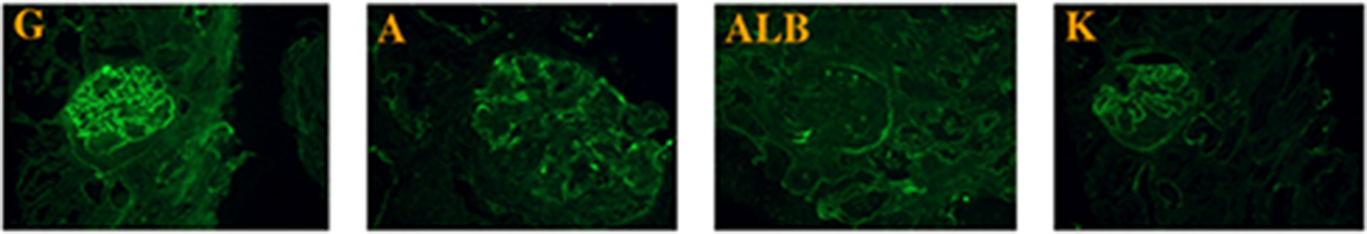
The presence of karyomegalic cells in the renal graft has many differential diagnoses. They present as KIN, BK nephropathy, or T-cell-mediated rejection. In KIN, the nuclei are large, pleomorphic, hyperchromatic, and diffuse in both proximal and distal tubules across the biopsy without intranuclear inclusion and tubulitis. In BK nephropathy, the nuclei are not pleomorphic and contain intranuclear inclusion bodies with focal distribution across proximal and distal tubules and tubulitis. In addition, viral stains, like SV40, are positive. In cell-mediated rejection, the nuclei are not pleomorphic or hyperchromatic and are present in areas of tubulitis without intranuclear inclusion bodies.
The occurrence of KIN in the renal allograft may be due to recurrence, donor-associated disease, or de novo. One of the limitations of this case report is the absence of genetic studies on two brothers, the unavailability of their native biopsies, and the non-performance of SV40 to exclude the diagnosis of BK nephropathy.


留言 (0)