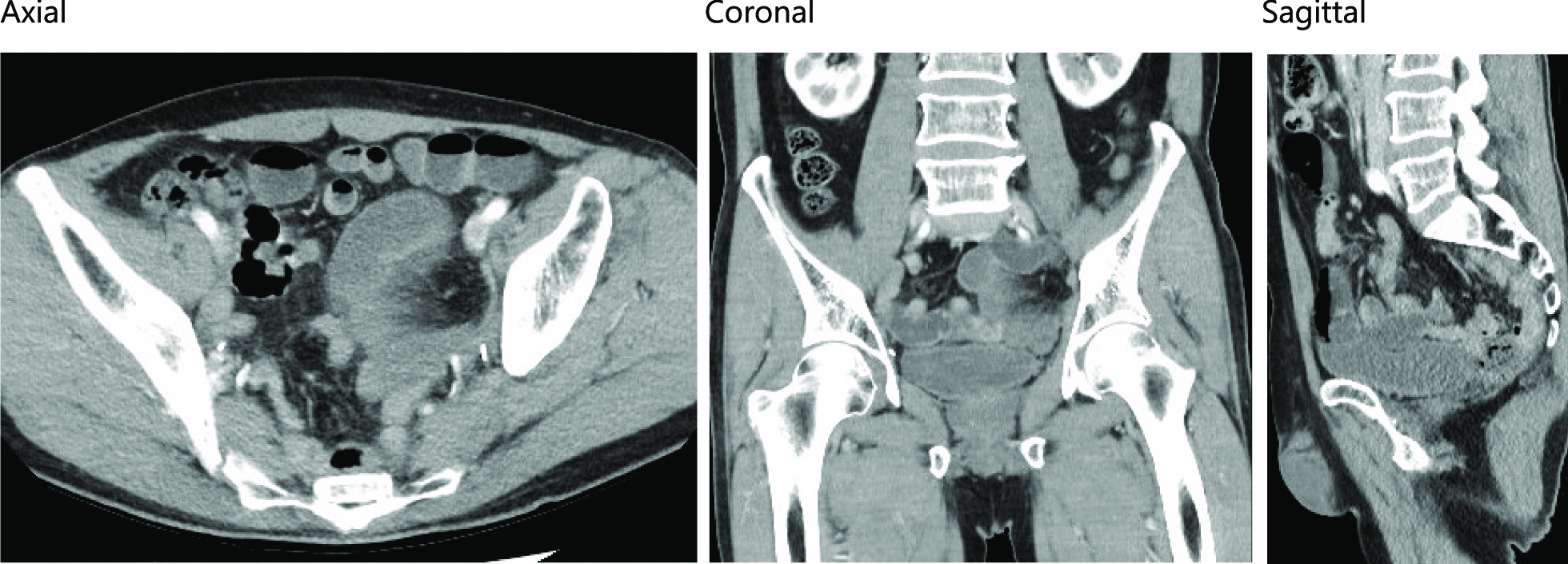
ILC is the most common of the special types of breast cancer. ILC accounts for up to 15% of all breast cancer. It has a unique metastatic presentation, with a predilection for common sites such as the liver, lung, and bone as well as the gastrointestinal tract and gynecologic organs. ILC is also characterized by the lack of E-cadherin, a tumor suppressor that plays an important role in epithelial cell–cell adhesion and tumor morphogenesis. Loss of E-cadherin expression leads to decreased cellular adhesion, resulting in cell migration and metastatic spread [2, 4, 5]. These distinctive features of ILC presumably led to the bone-marrow metastasis described in the current report. Borst et al. reported that the metastatic pattern of ILC differs from that of IDC. The rate of bone-marrow metastasis was 21.2% among 359 patients with ILC and 14.4% among 2,246 patients with IDC, a statistically significant difference [6]. The prognosis of bone-marrow metastasis is so poor that chemotherapy should be considered. We began treatment of our patient with paclitaxel plus bevacizumab. Her disease status improved sufficiently. Of note, initial therapy for metastatic breast cancer with paclitaxel plus bevacizumab was previously shown to prolong progression-free survival [7].
In the present case, the primary ILC lesion in the breast was only revealed with MRI based on characteristic NME. As ILC invades the adjacent breast tissue along mammary ducts and usually does not form a palpable lump, conventional mammographic and ultrasound challenges result in false-negative diagnoses [8]. Brem et al. reported that the sensitivity of mammography, US and MRI in the detection of ILC among 28 biopsy-proven cases were 79%, 68% and 83%, respectively, indicating that the MRI presented the highest sensitivity [9]. In the fifth edition of the American College of Radiology Breast Imaging Reporting and Data System (BI-RADS) lexicon, NME is classified as homogeneous, heterogeneous, clumped, and in a clustered ring [10]. NME lesions are considered to indicate malignancy; however, the prevalence of NME is much lower than that of mass enhancement [11, 12]. The BI-RADS lexicon has been shown to be inadequate for distinguishing between benign and malignant NME lesions [13,14,15]. In an analysis of 99 benign and 30 malignant NME lesions, Aydin reported that 17 (13.2%) lesions had a segmental distribution, like our patient’s lesion, of which 5 were benign and 12 were malignant [11]. Segmentally distributed lesions are most likely to be malignant.
In our case, second-look US failed to visualize the primary lesion in the breast that was detected by MRI. RVS was adopted as the next modality, which successfully demonstrated the lesion as a heterogeneous mass with low echogenicity and an irregular edge. It was previously reported that second-look US of breast cancer lesions detected with MRI identified 49% of mass lesions, 42% of focal lesions, and 15% of NME lesions [16]. RVS can be effective for clear visualization of NME lesions. The RVS system employed in this case simultaneously displayed both sonographic and MRI cutaway images of the same site in real time, which has excellent accuracy for identifying breast lesions with enhancement on MRI [3]. Nakano et al. also reported that RVS was highly useful for demonstrating NME lesions detected with MRI in 12 patients with breast cancer who underwent breast-conserving surgery [17].
For pathological confirmation of the lesions visualized only by MRI, MRI-guided biopsy has become increasingly available. However, these techniques are not commonly available and require the costly use of the device and the medical staffs. On the other hand, RVS does not require large equipment and can be carried out easily at any time, suggesting that RVS is a patient-friendly technique that can visualize a large proportion of MRI findings. Furthermore, it should be validated that RVS could select the patients that really undergo MRI-guided biopsy. Nakano et al. reported that the detection rates of MRI-detected lesions with second-look sonography using RVS or not were 90% and 30%, respectively (P < 0.001) [18]. There would be thoughts that the MRI-guided biopsy is applied for the lesions that RVS failed to visualize. Unfortunately, no direct comparison between MRI-guided and RVS-guided biopsy has been demonstrated. The comparison of vacuum-assisted biopsy (VAB) among MRI, stereotactically and ultrasound-guided was reported by Imschweiler et al. [19]. In that report, the technical success rates of MRI-guided, stereotactically guided and ultrasound-guided VAB were 98.4%, 99.1% and 99.6%, respectively. There was a significant difference in the technical success rates between the MRI and ultrasound-guided VAB (P < 0.001). The complications were hemorrhage, infection, lesion miss and so forth. The total complication rate of ultrasound-guided VAB was significantly lower than that of MRI-guided VAB (P < 0.001).
RVS technique have some limitations. First, the MRI data introduced into RVS system are constructed with supine-position MRI examination, which is not established method for diagnosing breast cancer. Second, the US examinations are considered to be poor at reproducibility, because the breast will change its shape easily and the quality of the examination depend on the examiner’s skill. The result of multi-institutional study which was conducted for further investigation of the role of RVS and the examination of the interinstitutional reproducibility is awaited.


留言 (0)