
記住我


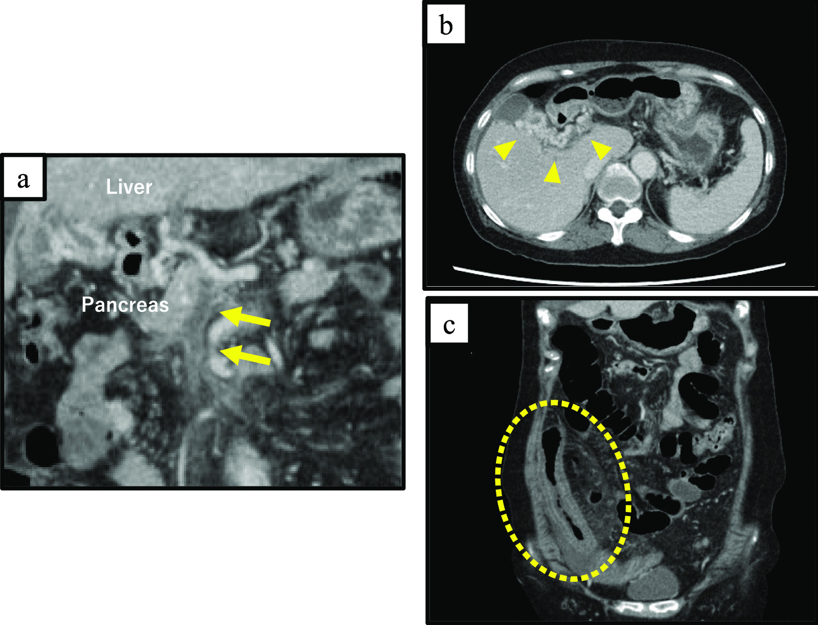

A man in his 80 s, with hematuria and edema of the right lower extremity had been hospitalized 2 months earlier and received medications for right sciatica. However, the symptoms exacerbated over time, and computed tomography (CT) revealed a 28-mm right IIAA and perianeurysmal fluid retention causing right pyelectasis. He was transferred to our hospital, on an emergency basis with a suspected ruptured IIAA. AAA was also detected, and although the IIAA appeared to have ruptured, the IIA was occluded (Fig. 1A, B). His medical history included pacemaker placement for sick sinus syndrome 22 years ago, percutaneous coronary intervention for angina pectoris 4 years ago, and aortic arch replacement for thoracic aortic aneurysm 1 year ago. At the time of hospitalization, his body temperature, blood pressure, and heart rate were 35.4 °C, 86/58 mmHg, and 61 beats/min, respectively. He showed gross hematuria and prominent edema of the right lower extremity. Blood tests revealed severe inflammation (white blood cell [WBC] count, 12,340/μL; C-reactive protein [CRP], 21.0 mg/dL), anemia (hemoglobin, of 9.9 g/dL), poor nutrition (serum albumin, 1.8 g/dL), and chronic kidney disease (blood urea nitrogen, 41.8 mg/dL; serum creatinine, 1.97 mg/dL). Systemic inflammatory response syndrome developed, and the infectious sources were assessed. Urinalysis revealed turbidity, positive occult blood, and increased WBC count. Through blood culture, Staphylococcus hominis was detected in one of four sets. Urine culture revealed the presence of methicillin-resistant Staphylococcus aureus and Enterococcus faecalis. Echocardiography demonstrated a 15-mm mass in the right atrium with movement at the pacemaker lead (Fig. 1C). The patient was diagnosed with a suspected secondary infected right IIAA related to urinary tract or pacemaker lead infection based on the above test results. Because the patient was strongly suspected of septic shock and not ruptured IIAA, systemic infection control to some extent was planned before radical treatment of the suspected infected aneurysm.
Fig. 1
A Preoperative computed tomography (CT) revealed a 28-mm right internal iliac artery aneurysm (IIAA, arrow) and perianeurysmal fluid retention (arrowheads). B Three-dimensional CT angiography showed abdominal aortic aneurysm (white arrow) and occluded IIA (black arrow). C Mobile 15-mm mass was observed at the tip of the pacemaker lead as a source of infection. D CT revealed increased perianeurysmal fluid retention on day 18 of hospitalization (arrowheads). E Aggravated hematuria with increased perianeurysmal fluid retention. F Endovascular aortic aneurysm repair (arrowhead) and coil embolization (arrow)
Ureterography revealed no evidence of communication between iliac arteries and right ureter and a ureteral stent was placed on the day of admission. Furthermore, the intravenous antibiotic administration of meropenem and vancomycin was initiated for treating urinary tract infection and pacemaker lead infection. On day 2 of hospitalization, the pacemaker was removed. On day 3, improvements in the WBC count (7030/μL) and CRP level (9.97 mg/dL) were observed following the above interventions. Therefore, systemic antibiotic dosing was planned for approximately 3 weeks before aneurysm treatment. However, low blood pressure was noted on day 18. CT revealed an increase in retroperitoneal fluid retention around the right IIAA, raising a suspicion of ruptured aneurysm and uretero-iliac artery fistula (Fig. 1D). Anemia and aggravated hematuria were also detected (Fig. 1E). Therefore, emergency coil embolization of the right IIA and endovascular aneurysm repair (EVAR) from the right common iliac artery to the external iliac artery (GORE®EXCLUDER®, Gore W L & Associates Inc., DE, USA) were performed to control bleeding and as diagnostic treatment on the same day (Fig. 1F). CT angiography and digital subtraction angiography showed no evidence of contrast enhancement of right ureter and urinary bladder; however, delayed retrograde arterial flow from distal branches of IIA might be associated with increase in fluid retention. Ureterography was not performed as the ureteral stent was already placed.
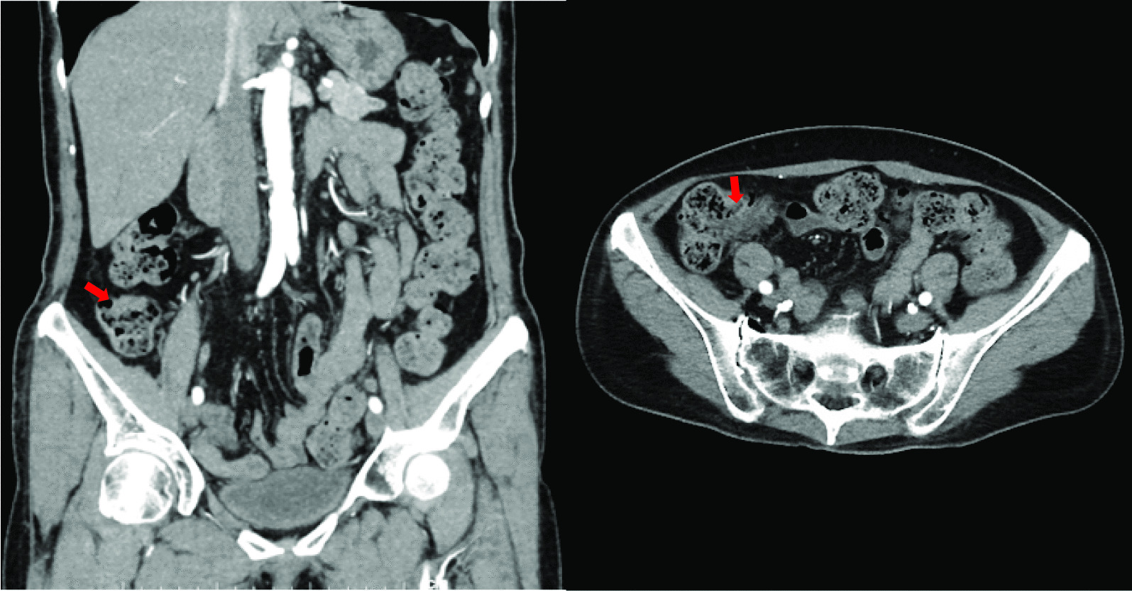
Subsequently, 2-[fluorine-18]-fluoro-2-deoxy-D-glucose (FDG) positron emission tomography (PET)–CT performed on day 21 revealed the accumulation of FDG, consistent with retroperitoneal fluid retention around the aneurysm (Fig. 2A). Based on his medical history and FDG PET–CT data, the patient was diagnosed with an infected aneurysm. EVAR followed by surgical abscess drainage and aneurysmectomy/vascular graft replacement at 1-week interval were scheduled as a two-staged treatment. The proximal part of the abdominal aorta was encircled with Teflon tape following median laparotomy, and the retroperitoneum around the right IIA was incised. No apparent perianeurysmal abscesses were found; however, a mass lesion in the iliopsoas muscle was detected. After this mass was incised, a cottage cheese-like white discharge was identified (Fig. 2B). The contents had serum components and bacterial culture of the components was negative. Intraoperatively, an iliopsoas abscess was clinically diagnosed, and only laparotomy irrigation drainage was performed with drainage tube replacement. However, the patient’s blood test results following the surgery revealed a strong inflammatory response and poor overall condition. Hence, on day 55, gallium scintigraphy was performed to assess residual abscesses, which revealed gallium-67 accumulation along the abscess cavity and ureters. Because the retroperitoneal infection was suspected to have spread to the ureter, the abscesses, ureter, and surrounding tissues were completely debrided (Fig. 2C). Abscess drainage and right nephroureterectomy were performed as the second laparotomy in collaboration with the urology department on day 79. On macroscopic examination, the distal third portion of the ureter was found to be enlarged. An incision was made to the enlarged ureter after nephroureterectomy; no purulent discharge was observed in the lumen, and only wall thickening was noted (Fig. 2D). Following the complete irrigation of the abdominal cavity and extensive debridement of tissues around the kidney and ureter, periarterial tissues were visually examined for infection, and the abdomen was closed as usual. After the second surgery, the pelvic lesion grew despite EVAR and coil embolization having completely controlled blood flow in the left IIA (Fig. 2E, F).
Fig. 2
A Positron emission tomography–computed tomography (CT) revealed accumulation consistent with retroperitoneal fluid retention around the aneurysm. B Intraoperative image of the first laparotomy. The iliopsoas muscle (arrows) had a white cottage cheese-like secretion inside. C, D Intraoperative image of the second laparotomy. The white necrotic tissues were resected to control infection. The right kidney and ureter were also resected. The distal part of the ureter was enlarged (thin black arrows). No purulent discharge was observed in the lumen, and only wall thickening (thick white arrows) was noted. E Postoperative CT angiography after the second laparotomy revealed complete isolation of blood flow in the IIA by endovascular aortic aneurysm repair (EVAR, arrowhead). F Pelvic mass grew despite EVAR (arrowheads)
The removed tissues were examined pathologically, which led to the definitive diagnosis of DLBCL (Fig. 3). Following consultation with the hematology department, we agreed that the patient’s poor overall state would make chemotherapy intolerable. The patient’s condition failed to improve, and he died on day 95 of hospitalization owing to multiple organ failure.
Fig. 3
Hematoxylin and eosin (HE) staining, immunohistochemical staining, and Epstein–Barr virus-encoded small RNA (EBER) in situ hybridization (ISH) in the resected perianeurysmal tissues. A HE staining (4 ×) showed diffuse growth of atypical cells with necrosis (upper left). B HE staining (40 ×) showed medium-to-large atypical lymphoid cells infiltrated in the tissue, suggesting diffuse large B-cell lymphoma. C Atypical lymphoid cells were positive for CD20, the classic marker of B-cells (40 ×). D EBER–ISH showed positivity in the neoplastic cells (40 ×)
留言 (0)