Proximal humeral fracture is a common osteoporotic fracture in the elderly. It usually occurs in low-energy fragility fractures. With the advent of global aging, the incidence of proximal humeral fracture is increasing every year [14, 15]. However, the treatment of proximal humeral fracture with the comminuted medial wall remains controversial because of the high incidence of postoperative complications [1, 16]. The integrity of the medial wall of the proximal humerus plays an important role in its postoperative stability and its biomechanical properties are similar to those of the proximal femur [12, 17]. At present, it has been accepted that comminuted fracture of the medial calcar with the bone defect may result in the varus of the humeral head, which is the most common pattern of internal fixation failure [18]. ORIF using PHILOS plate (Synthes) is still the most commonly used surgical method for the treatment of proximal humeral fractures [19]. To prevent varus deviation of the humeral head, the medial calcar screws must be inserted along the inferior tangential direction of the medial cortex to enhance fracture stability during the treatment of a injured medial calcar. However, in some patients, the medial cortex is severely disrupted and the calcar continuity cannot be maintained after reduction; or in the case of extremely serious osteoporosis, varus deformity of the humeral head may occur even after fixation with the calcar screws. In view of the high incidence of postoperative complications, some investigators have suggested that an adjuvant plate or fibular strutallograft (FSA) could be used for additional fixation in addition to PHILOS [20, 21]. The plates can be placed in various positions, including the anterior and medial to the proximal humerus [6, 22]. However, the technique of anterior plate assisted internal fixation has rarely been described, and few studies have been performed on the effect of lesser tuberosity fractures and their associated medial calcar disruption on patients with proximal humerus fractures.
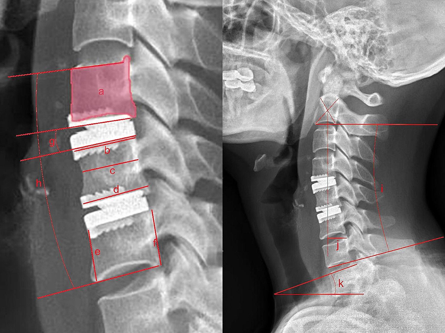
In recent studies, researchers have begun to pay attention to the role of the lesser tuberosity in the stability of the proximal humerus. Katthagen JC et al. conducted a biomechanical study on whether the 4-part proximal humeral fractures were stabled by the lesser tuberosity fixation. The results have shown that the use of screw-assisted fixation in the 4-part proximal humeral fractures was effective in improving the stability of the proximal humerus [9]. This study also demonstrated the key of the lesser tuberosity on the structural stability of the proximal humerus. In our study, a correlation was found between the comminution of the lesser tuberosity (fragments > 2) and the destruction of medial calcar. The number of patients in Group I and Group IV was 98, which was significantly higher than in Group II and Group III. Considering that the medial aspect of the lesser tuberosity is connected with the humeral medial calcar, we believe that if the lesser tuberosity is comminuted and displaced on X-ray, it indicates the destruction of the calcar. In addition, when the calcar disruption, the change of the neck-shaft angle was 9.37° ± 3.53° for an intact lesser tuberosity and 13.48° ± 8.91° for a comminuted lesser tuberosity. Therefore, the fragmentation of the lesser tuberosity further predicts the occurrence of postoperative humeral head collapse, which may be related to the following reasons. When there is lesser tuberosity comminution and humeral medial calcar injury, the patient’s fracture is severely displaced, and the anterior varus of the humeral head often occurs. At this point, the reduction becomes more difficult and there is a lack of support for the medial side of the humerus after the operation. However, the free tuberosity fragment was not fixed intraoperatively and the proximal humerus was unsupported anteriorly. Besides, patients with preoperative osteoporosis have relatively slow postoperative functional recovery, which further aggravates the osteoporosis, and may lead to inadequate s postoperative screw support.
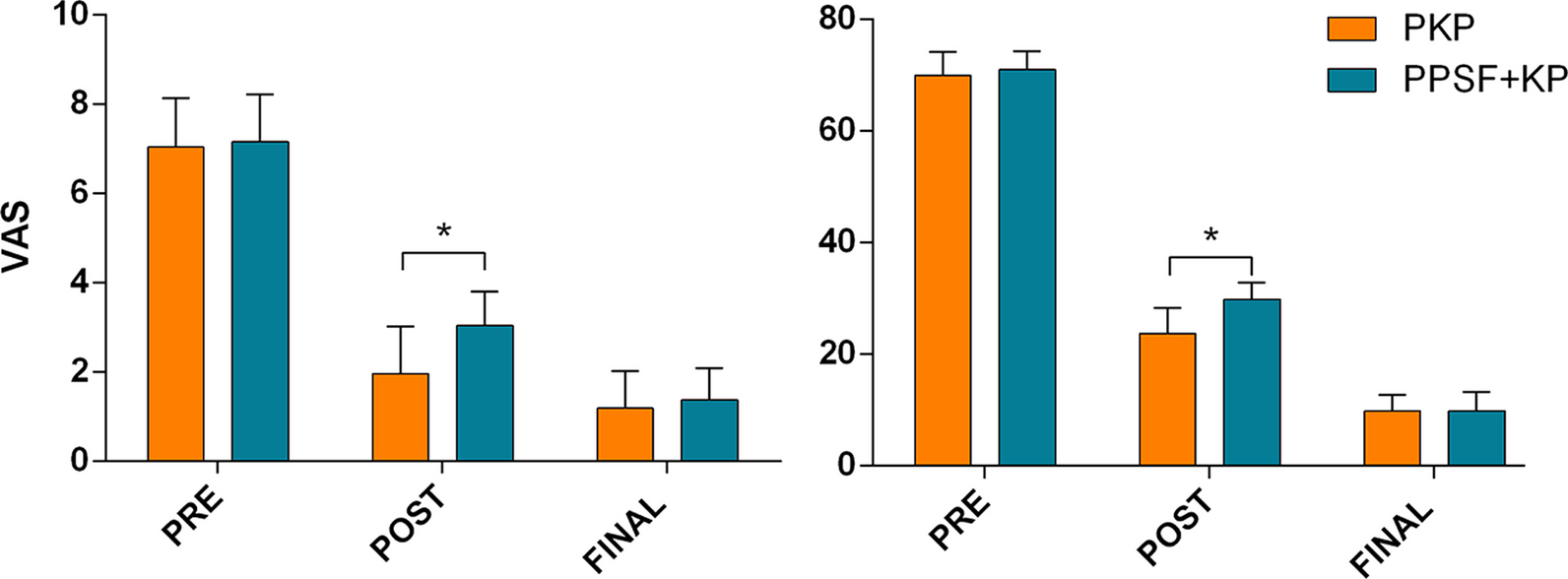
In our study, although there was no difference in VAS scores between groups 1 year after the operation, the results of DASH upper limb function scores were similar to the variations of humeral neck-shaft angle. It was believed that this was due to the functional impact of the humeral head collapse. Furthermore, regarding the fact that the patient's lesser tuberosity comminution may lead to the injury of subscapular muscle, the positive rate of lift-off test 1 year after surgery was analyzed. As expected, the positive rate of lift-off test was higher in Group III and Group IV patients 1 year after the operation, indicating that lesser tuberosity comminution may affect the function of the subscapularis muscle, which needs further research and analysis. If the function of the subscapularis muscle is affected, it will not only lead to the loss of shoulder adduction function but also further lifting and pronation function. The unsatisfactory reduction of the fracture at the lesser tuberosity may affect the matching of the humeral head and the shoulder glenoid cavity, resulting in incorrect trajectory of the humeral head over the shoulder glenoid cavity (similar to the off-track of Hill-Sachs injury) [23], and even the possibility of postoperative shoulder instability. Therefore, it is not sufficient to rely on the LLP solely to treat medial calcar disruption accompanied by lesser tuberosity comminution. The medial bone grafting combined with steel cable can be used to bind and fix the fractures in some patients. However, it may not able to completely tie the severely fragmented anterior lesser tuberosity fragments, and the anterior plate is required to assist in fixation.
The study has some limitations. First, it is a retrospective study in which some patients with relatively severe fractures of the lesser tuberosity opted for conservative treatment, while other patients with severe fractures of the lesser tuberosity and medial calcar were treated with shoulder arthroplasty, which may have altered the proportion of patients in the different groups. In addition, the range of motion of the shoulder joint was not evaluated in all patients. Future prospective investigations should clarify the results of this study.


留言 (0)