
記住我


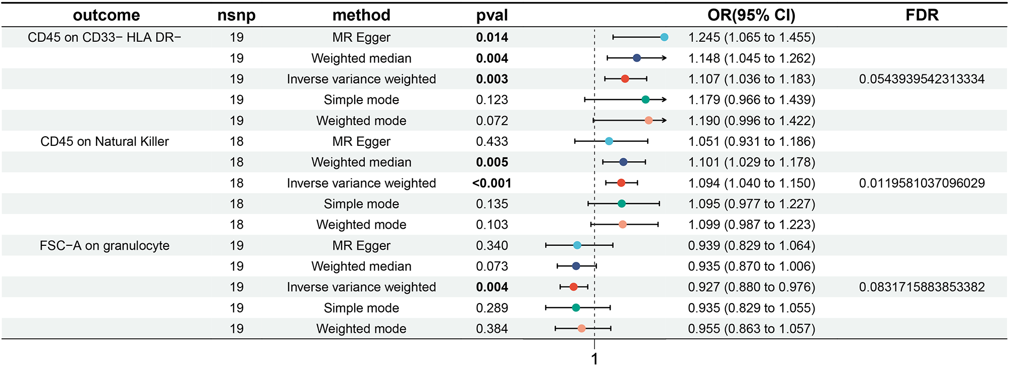

A 33-year-old previously healthy woman presented with bloody diarrhea and nausea. An ECG was performed as part of the routine procedures of the emergency room. The electrocardiogram revealed a myocardial infarction with ST segment elevation in leads II, III, aVF and inverted T-waves in leads V1-V3 (Figs. 1, 2) as well as a flattening of T-waves in V7-V8 and inverted T-waves in V9 (Fig. 3). The patient did not state to have any chest pain.
Fig. 1
Electrocardiogram, standard limb leads, showing ST segment elevation in leads II, III, aVF
Fig. 2
Electrocardiogram, chest leads, showing inverted T-waves in leads V1-V3
Fig. 3
Electrocardiogram, chest leads, showing flattening of T-waves in leads V7-V8 and inverted T-waves in V9
She was not taking any medication. On initial evaluation, her blood pressure was 94/76 mmHg, heart rate was 98 bpm. Upon cardiac auscultation, there were no audible murmurs, and her lungs were clear. No oedema and no signs of congestion were noted. There was slight tenderness on palpation throughout the abdomen.
Laboratory data showed a significant increase of high-sensitivity cardiac troponin-T (578 pg/mL; normal: < 14 pg/mL), creatine kinase (317 U/L; normal: < 167 U/L), creatine kinase-MB (52 U/L; normal: < 24 U/L), C-reactive protein (191 mg/L; normal: 0–5 mg/L), lactate dehydrogenase (236 U/L; normal: 135–214 U/L) and D-dimer (16.95 µg/mL; normal: < 0.5 µg/mL). Other abnormal laboratory data included a decreased platelet count of (85 G/L; normal: 150 – 370 G/L).
Hemoglobin (14 g/dL; normal: 12 – 15.6 g/dL) as well as coagulation factors showed no abnormality. Further laboratory results, including lipids, glucose and electrolytes were also within the normal range.
An urgent coronary angiography revealed a thrombotic occlusion of the periphery of the left circumflex coronary artery (Video 1). Pre-treatment with acetylsalicylic acid was not given, however the patient received 5000 IU heparin. Due to the localization of the thrombus in the vascular periphery, stent implantation was not feasible. Probing with a wire and a non-inflated percutaneous coronary intervention balloon was performed, but distal perfusion was not restored. The patient received a bolus of Tirofiban, however no permanent infusion of the medication was given due to the persisting intestinal bleeding. Dual antiplatelet therapy with acetylsalycylic acid 100 mg and ticagrelor 90 mg b.i.d. was initiated according to the ESC guidelines [9].
The echocardiographic examination revealed a normal left ventricular ejection fraction (Video 2). No segmental ventricular wall motion abnormalities and no ventricular thrombus were observed. In the transesophageal echocardiography (TEE) no intracardiac thrombus and no structural anomaly could be detected. Due to low imaging quality of the first study, we repeated the TEE study. Neither a thrombus nor a patent foramen ovale, also using a bubble test, could be detected in the additional transesophageal echocardiography.
A cardiac magnetic resonance imaging (MRI) was carried out. There was transmural late gadolinium enhancement (LGE) of the basal and mid inferior left ventricular segments, corresponding to an ischaemic distribution pattern, as well as increased signal intensity of the basal and mid inferior left ventricular segments (Figs. 4, 5, 6 and 7). Cine sequences revealed a moderately reduced LV systolic function with hypokinesis of the inferior myocardial segments and an LVEF of 46% (Video 3).There was no evidence of a left ventricular thrombus. While no abnormalities were detected in echocardiography, cardiac MRI showed regional wall motion abnormalities, probably due to better definition of the endocardium as well as limited echocardiographic assessability due to tachycardia during echocardiography.
Fig. 4
Cardiac MRI, inverse recovery LGE sequence, long axis view, showing transmural late gadolinium enhancement of the mid inferior segment in an ischemic distribution pattern
Fig. 5
Cardiac MRI inverse recovery LGE sequence, midventricular short-axis view, showing transmural late gadolinium enhancement of the mid inferior segment in an ischemic distribution pattern
Fig. 6
Cardiac MRI, T2-weighted stir sequence, long-axis view, showing an increased signal intensity of the mid inferior and basal inferior segment
Fig. 7
Cardiac MRI, T2-weighted stir sequence, midventricular short-axis view, showing an increased signal intensity of the mid inferior segment
Further diagnostic work up identified a highly active ulcerative pancolitis (Fig. 8). An infectious cause of the dysentery was ruled out. Hepatitis, human immunodeficiency virus, cytomegaly virus and tuberculosis were ruled out.
Fig. 8
Colonoscopy showing mucosal ulcerations extending into deeper layers and loss of vascular pattern
An extensive diagnostic work up for cardiac thromboembolism was performed [10].
Although we found evidence of vascular thrombosis on the coronary angiogram, there was no history of pregnancy morbidity and the patient tested negative for antiphospholipid antibodies, thus the revised Sapporo Criteria for Antiphospholipid Syndrome (APS) were not met, rendering a primary antiphospholipid syndrome as cause of the myocardial infarction very improbable [11]. Clinical criteria for Systemic Lupus Erythematosus (SLE) were absent, and there was a negative test result for Antinuclear Antibodies (ANA), also eliminating an SLE-associated antiphospholipid syndrome as an explanation [11].
A test for factor V Leiden mutation or a Factor II-20210A mutation was negative, thus these types of hereditary hypercoagulability were not taken into consideration as a differential cause for the myocardial infarction [10].
In the absence of clinical clues or coronary malformations, mid-size vessel vasculitis as a cause of the myocardial infarction was highly unlikely. In addition, there were negative results for Anti-neutrophil cytoplasmic antibodies (ANCAs), making an ANCA-associated vasculitis as a cause of the myocardial infarction even more improbable [12].
A coagulopathy was also conceivable in the context of a COVID-19 infection [13]. We ruled this possibility out through a PCR Test.
Atrial fibrillation as cause [10] of cardiac embolism was not recorded in a 7-day clinical monitoring period.
Potentially thrombogenic medication was not present. The patient was not taking oral contraceptives or anti-inflammatory drugs [14].
Thromboembolism in the context of acute bleeding was suggestive of disseminated intravascular coagulation (DIC). The ISTH Criteria for Disseminated Intravascular Coagulation [15] were not suggestive of overt DIC but a non-overt DIC could not be ruled out.
Inflammatory bowel disease (IBD) was first diagnosed in our patient and she had been without treatment.
Inflammatory markers were strongly elevated (C-reactive protein 191 mg/L; [normal: 0–5 mg/L]). We considered this to be an expression of an acute flare of the ulcerative colitis. The patient was treated with prednisolone 60 mg once daily and mesalazine 3 g once daily. Because of the lack of macroscopic improvement, we also initiated immune-modulating therapy with infliximab 300 mg I.V. Although the use of corticosteroids may be a risk factor for mechanical complications of myocardial infarction [16], we considered their use mandatory because of the high inflammatory IBD activity.
The developement was complicated by difficult-to-control lower gastrointestinal bleeding with a decreased hemoglobin value of 5.6 g/dL in the setting of ulcerative colitis and dual antiplatelet therapy with acetylsalycylic acid 100 mg and ticagrelor 90 mg b.i.d. The patient received two blood transfusions and the antithrombotic therapy was initially switched to to acetylsalycylic acid 100 mg and enoxaparin 60 mg once daily, but only after administration of infliximab and further modification of anticoagulative and anti-aggregatory therapy to apixaban 2,5 mg b.i.d. the lower gastrointestinal bleeding stopped.
On the day of discharge, the patient stated to now experience chest pain. A pericardial effusion was seen on the subsequent echocardiogram. We interpreted this in the context of Dressler syndrome and administered a therapy with colchicine for three months. Furthermore, we suggested to the patient that she should visit a specialized coagulation outpatient clinic. We scheduled a follow up with an ambulant care physician where she received the second dose of infliximab 300 mg I.V. two weeks later.
留言 (0)