
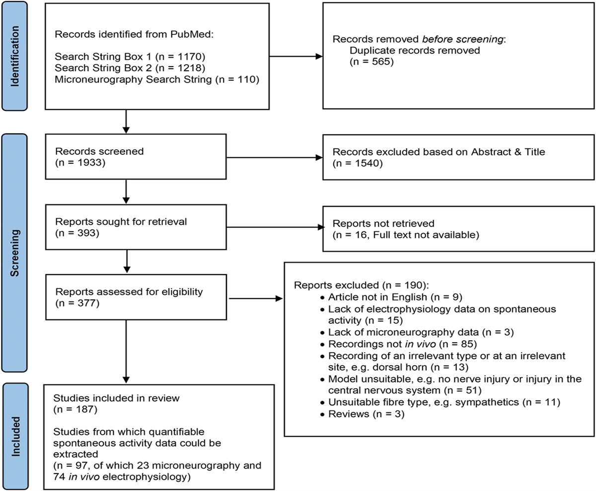
記住我


Tissue inflammation or injury increases the excitability of primary afferent neurons,10,16,17,25,28,44,70,72 amplifying their input toward the nociceptive central nervous system (CNS) networks and resulting in enhanced pain sensitivity, namely, hyperalgesia.4,25,78 The “first stations” processing the inputs from primary afferents are the intricate neuronal networks of superficial laminae of the dorsal spinal cord (SDH).6 These pain-related neuronal networks are composed of local excitatory and inhibitory neurons converging into projection neurons, which relay the information to higher brain centers, thus forming the sensation and perception of pain.11,75,76 It is well documented that inflammation and injury-mediated changes in the primary afferent input lead to increased excitatory synaptic transmission toward local interneurons and projection neurons.1,46,58,69 In other neuronal systems, enhanced synaptic bombardment and increased neuronal activity trigger an adaptive decrease in intrinsic neuronal excitability, hence regulating neuronal output.26,41,56,57 The adaptive changes tuning the activity of pain-related SDH neurons, thus controlling nociception and pain, have not been described before.
It has been demonstrated that the plasticity of axon initial segment (AIS) is an essential mechanism for tuning neuronal and hence network output in response to changes in input.12,27,38,47,49 The AIS is located at the proximal axon and harbors a dense array of voltage-gated channels and a unique repertoire of submembranous cytoskeletal scaffolding proteins, such as ankyrin G, clustering these channels together.27,31,38 The high density of voltage-gated channels allows AIS to convert graded synaptic inputs into a zero-one binary output of action potential (AP) firing. Notably, the size and position of individual AIS are dynamic and modified in response to physiological and pathological changes in neuronal activity.23,26,41,42,52,55,56 This structural AIS plasticity affects neuronal excitability, thus fine tuning neuronal output and the overall neuronal network response to changes in input.26,41,52,57,67 Sensory enrichment or pharmacologically induced increase in neuronal activity, for example, produces a distal shift of AIS location and AIS shortening, leading to a decrease in neuronal excitability.26,41,56,57 However, it is unknown whether inflammation or tissue injury–induced increase in input to SDH neurons induces plasticity of AIS, thus affecting the excitability of SDH neurons.
We studied the changes in the location of AIS in inhibitory and excitatory SDH neurons following hyperalgesic inflammation and detailed how these changes alter the intrinsic neuronal excitability. We show an inflammation-mediated distal shift of the AIS away from the soma in inhibitory but not excitatory SDH neurons, concomitant with the peak of inflammatory pain. This AIS translocation was accompanied by a decrease in the excitability of the inhibitory neurons. Following recovery from inflammatory hyperalgesia, the AIS location and neuronal excitability reversed to baseline levels. The computational model of SDH inhibitory neurons predicts that the distal shift of AIS is sufficient to decrease the intrinsic excitability of these neurons. The inflammation-mediated distal shift of AIS and the resulting decrease in inhibitory neurons' excitability could contribute to an increase in output from the pain-related SDH network, thus facilitating inflammatory pain.
2. Methods 2.1. AnimalsAll procedures were performed in accordance with the guidelines of the Animal Ethics Committee of the Hebrew University of Jerusalem and were approved by the Ethic committee of The Hebrew University. Five- to 7-week-old male Sprague Dawley (SD) rats (150-225 g) were housed under controlled temperature (23 ± 2°C) and environment, with ad libitum access to food and water, and kept in a 12-hour light/dark cycle. Our preliminary results from 10 female rats show poor AIS staining in spinal cord slices such that we could not detect spinal cord neurons expressing both Pax2 and ankyrin-G (see below, Immunohistochemistry section). Therefore, we continued our experiments using only male rats.
2.2. Spinal cord slices preparationSpinal cord slice preparation was performed as previously described by Lu et al.61 In short, rats were anesthetized with isoflurane and decapitated. The vertebral column was quickly removed and immersed in an ice-cold dissecting solution (in millimolar): 87 NaCl, 2.5 KCl, 1.25 NaH2PO4, 26 NaHCO3, 6 MgCl2, 0.5 CaCl2, 20 glucose, 77 sucrose, 1 kynurenic acid, oxygenated with 95/5% O2/CO2. After nerve roots and meninges were gently removed, the lumbar spinal cord was embedded in low melting agarose (3% in dissecting solution), and parasagittal slices of 300 µm were obtained. Spinal cord slices were then immersed in an oxygenated recovery solution (same as dissecting solution but without kynurenic acid) at 35°C and allowed to recover for 45 minutes. After 45 minutes, the slices were transferred to an oxygenated storage solution (same as recovery solution but at room temperature) and until recording.
2.3. ElectrophysiologyAfter recovery, slices were transferred to a submersion recording chamber and mounted on the stage of an upright microscope (BX51WI; Olympus Corporation, Tokyo, Japan). Slices were then perfused with artificial cerebrospinal fluid (aCSF) containing the following (in millimolar): 125 NaCl, 2.5 KCl, 26 NaHCO3, 1.25 NaH2PO4, and 20 glucose oxygenated with 95/5% O2/CO2.
The internal solution was as follows (in millimolar): 130 K-gluconate, 6 KCl, 8 NaCl, 2 MgATP, and 10 HEPES (pH = 7.4 with KOH). The membrane potential was corrected for a liquid junction potential of +14.9 mV.
Whole-cell current-clamp recordings were performed from neurons located within lamina II, which was identified as a translucent band across the dorsal horn under the microscope. Voltages were recorded in fast current-clamp mode using a Multiclamp 700B amplifier (Molecular Devices , San Jose, CA) at room temperature. Data were low-pass filtered at 10 kHz and sampled at 20 kHz for 500-millisecond current steps protocols and 250 kHz for 10-millisecond current steps protocols (−3 dB, 8-pole Bessel filter). Patch pipettes were pulled using thick-walled borosilicate glass capillaries (1.5:0.86 mm, outer to the inner diameter) on a P-1000 puller (Sutter Instruments Co, Novato, CA) with a resistance of 3 to 5 MΩ when filled with standard internal solution (see above) and access resistance was maintained at 7 to 13 MΩ.
Command current protocols and data digitizing were generated with a Digidata 1440A A/D (Molecular Devices), interfaced with pCLAMP 10.3 (Molecular Devices). Data were analyzed using Clampfit 10.3 software and MATLAB.
For each analyzed SDH neuron, first, the firing profile (tonic or delayed) was determined as the patterns of the APs in response to 500-millisecond depolarizing square pulses (with increments of 0.01 nA). The resting membrane potential (RMP) was maintained at −70 mV in all the analyzed neurons to eliminate the effect of potassium IA current inactivation on the firing pattern.62 To ensure the stable firing pattern of the neurons, ie, that delayed-firing and tonically firing neurons keep their properties regardless of the injected current, all recorded neurons analyzed were classified following measurements at a wide range of current steps.
Apparent input resistance (Rin) was measured using a series of 500-millisecond hyperpolarizing and depolarizing square pulses (with increments of 0.01 nA). Rin was calculated as the slope of the voltage–current (V-I) curve in the linear part of the hyperpolarizing range.
The latency to the first AP was measured following a 500-millisecond depolarizing pulse of 150 pA, as the time between the beginning of the stimulation step to the peak of the first AP.
Single APs were evoked by 10-millisecond depolarizing current pulses of 0.01-nA increments. The threshold current (rheobase) was measured as the minimum current required to evoke an AP. The phase plots of dV/dt vs membrane voltage were formed by plotting the rate of change of membrane potential with respect to time (dV/dt) as a function of membrane potential, following a suprathreshold 10-millisecond current pulse. Maximum velocities (V/s) were obtained from (dV/dt)max. The AP thresholds were obtained by analyzing the phase plots (dV/dt) when plotted vs time and vs membrane voltage. The threshold voltage was detected using the “first local minimum,” which was determined by analyzing the function from its positive peak to time “0” as the first minimal value of dV/dt after the peak, followed by an additional increase in the dV/dt. Then, the time of the first local minimum was detected, and its voltage was assessed from the original trace and defined as the threshold voltage.
2.4. ImmunohistochemistryRats were deeply anesthetized with ketamine and xylazine and fixed by intracardiac perfusion with 20 mL of 4% paraformaldehyde (PFA) freshly prepared in phosphate-buffered saline (PBS, pH 7.4) at 4°C. The lumbar spinal cord was then immediately dissected out, followed by immersion for 1 hour in 4% PFA at 4°C, and cryoprotected by overnight immersion in 30% sucrose in PBS at 4°C. Tissue samples were then frozen in optimal cutting temperature (OCT) medium, and 30-μm cryosections were collected onto Superfrost Plus slides and stored at −20°C.
Slides were first thawed at room temperature (RT), washed in PBS, and incubated for 7 minutes in a permeabilization solution (0.5% Tween-20 and 1% Triton X-100 in PBS), followed by incubation of 1 hour with blocking solution (0.3% Triton X-100, and 5% donkey serum in tris-buffered saline [TBS]).
Sections were then incubated overnight at 4°C with primary antibodies, diluted in antibody solution (0.1% Tween-20 and 3% bovine serum albumin in PBS). Then the samples were washed 3 times for 10 minutes in PBS, followed by incubation in the dark with fluorescent secondary antibodies, diluted in antibody solution (0.1% Tween-20 and 3% bovine serum albumin in PBS) for 1 hour at room temperature. Finally, sections were washed 3 times for 10 minutes in PBS, then dried out, and mounted with Vectashield.
2.4.1. AntibodiesThe triple labeling was performed using (1) mouse monoclonal Ankyrin-G (1:250, NeuroMab Facility, Cat# 75-146) with Alexa Fluor 647-conjugated AffiniPure Fab Fragment Goat Anti-Mouse (1:1000, Jackson Immunoresearch, West Grove, PA, Cat# 115-607-186) as secondary antibody; (2) guinea pig polyclonal Anti-NeuN (1:500, Merck, Darmstadt, Germany, Cat# ABN90P) with Alexa Fluor 488-conjugated AffiniPure Donkey Anti-Guinea Pig (1:1000, Jackson Immunoresearch, Cat# 706-545-148) as secondary antibody; and (3) rabbit polyclonal anti-Pax2 (1:500, Invitrogen, Molecular Probes, Eugene, OR, Cat# 71-6000 with DyLight 405-conjugated AffiniPure Goat Anti-Rabbit (1:1000, Jackson Immunoresearch, Cat# 111-475-003) as secondary antibody.
2.4.2. Image analysisImages were acquired using a confocal microscope (Axiovert 200 M, Zeiss; Le Pecq, France) and a 60X objective. Images were processed with the NIH ImageJ software (Bethesda, MD). Immunofluorescence intensity profiles were obtained from a segmented line traced along the axon segment on a stack-of-interest projected image using ImageJ. Fluorescence intensity values were normalized to values between zero to one. The distance of the AIS from soma was determined while analyzing Alexa Fluor 647 as the point at which the fluorescence value reached 0.33. Each figure corresponds to a projection image from a z stack of 0.5-μm distant optical sections, limited to the object of interest. To minimize the measurement errors resulting from the z stack processing, we limited the number of maximum stacked sections to 6. Because of this limitation, the length of the AIS itself could not be analyzed because such analysis required much more than 6 sections. In these experiments, the yield of identifying excitatory or inhibitory neurons in male rats with labeled AIS was very low, and no more than one neuron per animal was analyzed, such that the numbers indicated in the figures correspond both to the number of neurons and the rats used in the experiments. In female rats, we could not detect neurons showing both labeling for Pax-2 and ankyrin-G (n = 10 rats), and therefore, we excluded them from the analysis.
2.5. Behavioral experiments 2.5.1. Paw withdrawal latencyThe paw withdrawal latency was measured using Hergeaves apparatus in both hind paws, as previously described.33 The rats were habituated to the test environment for 1 week in Plexiglas chambers and the experimental surfaces before the behavioral tests. The behavioral baseline was obtained by 3 preliminary measurements. The same investigator performed the scoring in all the behavior tests. Injected solutions were freshly prepared on the day of the experiment.
Animals received a 30-μL intraplantar injection of complete Freund adjuvant (CFA; Calbiochem EMD Chemicals, San Diego, CA) in the left hind paw. Twenty-four hours after CFA injection, the thermal threshold was measured. The behavioral tests were repeated on days 2, 3, 6, 9, 12, and 21.
2.6. Computer simulationThe simplified inhibitory interneuron model was implemented in NEURON 7.8 with the Python interpreter.19,36 We based our model on a previously published computational model of the SDH inhibitory neuron,63 with adjustments detailed below made to explore the effect of AIS plasticity on cell excitability.
2.6.1. MorphologyThe morphology of the tonically firing inhibitory neuron was based on Melnick et al63 and included a cylindrical soma (10 µm in diameter, 10 µm in length) connected to an equivalent dendrite segment. The dendrite was 500 µm in length and 0.86 µm in diameter. These parameters were derived from the neuronal properties as described below in “Calculation of dendrite length.” To study the effects of AIS distancing, we modeled an axon hillock, which served as a spacer between the soma and the AIS. Because the geometrical and physiological properties of the axon hillock in SDH inhibitory cells were not previously described, we modeled 4 possible axon hillocks configurations: (1) an “active” cone-shaped axon hillock, which decreased gradually from 3 μm in its proximal end to 1.3 μm in the distal end and included somatic active conductances described below; (2) a “passive” cone-shaped axon hillock, which decreased gradually from 3 μm in its proximal end to 1.3 μm in the distal end and included somatic passive conductances described below; (3) an “active” cylindrical axon hillock set to 1.3 μm in diameter; and (4) a “passive” cylindrical axon hillock set to 1.3 μm in diameter. The subsequent segment was the AIS compartment with a constant diameter of 1.3 μm, which terminated with a sealed-end condition. To simulate changes in AIS location, we gradually increased the length of the axon hillock from 1 to 25 μm while keeping all other aspects of neuron morphology and physiology constant. We used the AIS length of 25 μm as was suggested by Melnick et al.63 and because this length is an average of AIS length variations used in computational models studying AIS.24 To rule out the effect of AIS length on the AIS distancing-mediated changes on the threshold current, we also performed a series of stimulation with AIS of 15 μm.
2.6.2. Passive membrane propertiesA membrane capacitance of 1 μF cm−2 was set for all compartments.63 The axial resistance was set at 80 Ω cm.63 To prevent spontaneous firing of the cell following changes to axon initial segment morphology, we set the passive membrane resistance to 20,000 Ω cm2 (passive membrane conductance = 5 × 10⁻⁵ S cm−2) uniformly for all compartments. The equilibrium potential for passive conductance was −70 mV, and the resting membrane potential was −70 mV.63
2.6.3. Calculation of dendrite lengthFollowing the adjustment of specific passive resistance, the parameters of an equivalent dendrite were calculated using the procedure described previously.18,65,66 We used the electronic length of 0.68 and Rin of 1.7 GΩ adapted from the study of Melnick et al.63 We calculated dendrite length and diameter using the parameters described above applied to the equations describing a passive cable:λ2=DRm4RiRin=4λRiπD2cothL,l=Lλ,where Rm is a membrane resistance, Ri is an axial resistance, Rin is an input resistance, λ is a characteristic length, D is the diameter of the equivalent dendrite, and l is the equivalent dendritic length. Accordingly, the calculated dendrite dimensions were a length of 500 μm and a diameter of 0.86 μm.
2.6.4. Active conductancesThe model includes sodium (Na), and potassium delayed rectifier (KDR) currents as previously described,63 which were fit to Hodgkin–Huxley style equations. The steady-state activation variable (m∞) and the time constant of activation (τm) were determined as m∞ = αm/(αm + βm) and τm = 1/(αm + βm) adapted from the original model. For simulating the excitable properties of a neuron, we used the following fixed maximal conductance parameters for each of the segments, maintaining a higher active conductance density at the AIS compared with the remaining neuron.
(1) gKDR Dendrite = 0.034 S cm−2 (2) gKDR Soma = 0.0043 S cm−2 (3) gKDRAIS = 0.076 S cm−2 (4) gNa Soma = 0.008 S cm−2 (5) gNa AIS = 1.8 S cm−2.The reversal potentials for sodium (ENa) and potassium (EK) were set to +60 mV and −84 mV, respectively. The passive reversal potential was set to −70 mV. All conductances were evenly distributed across the model.
2.6.5. Stimulation parametersAll recordings were performed by positioning a NEURON “point-process” electrode at the center of the soma. For computational precision, all compartments were divided into many segments so that the length of individual segments was usually below 1 μm. All simulations were run with 0.05-μs time steps, and the nominal temperature of simulations was 23°C.
2.6.6. Data analysisData generation and plotting were performed with custom-written Python 2.7 code. To avoid artifacts resulting from the artificial instability of the NEURON simulation environment before reaching steady state, we allowed for a long 500-millisecond initialization time before stimulation of the simulated neuron, and this time was discarded from the analysis.
2.7. Statistical analysisStatistical analyses were performed using Prism 7 (GraphPad). Quantitative data were expressed as the mean ± SD. We did not perform a power analysis because the proposed experiments are novel, and therefore, we cannot estimate the effect size; however, sample sizes in this study were similar to previous reports.61 Rats were randomly allocated to groups in all experiments. All the analyzed data were normally distributed. The normality was assessed using the Shapiro–Wilk test. Two-tailed unpaired Student t-tests, ordinary 1-way analysis of variance, and contingency Fisher exact test were used when appropriate. Actual P values are presented for each data set. The criterion for statistical significance was P < 0.05. Boxplots presented in the figures depict the mean, 25th; 75th percentile, and SD.
2.8. Data and code availabilityAll data sets generated and/or analyzed during this study are available in the main text or upon request. The model files are uploaded to Model DB and are also available on request.
Further information and requests for resources and reagents should be directed to and will be fulfilled by the Lead Contact, Alexander Binshtok ([email protected]).
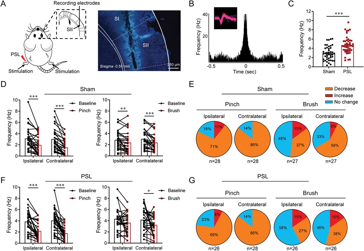
3. Results 3.1. Axon initial segment in inhibitory but not excitatory spinal cord dorsal horn neurons shifts away from the soma during inflammatory hyperalgesiaInflammation or tissue injury produces an increase in the gain of primary nociceptive neurons,5,10,25,39,54 thus enhancing their output toward the second-order spinal cord neurons.1,69 It has been demonstrated in various neuronal systems that abrupt changes in either intrinsic or synaptic activity lead to changes in the size and location of AIS, thus inducing adaptive changes in neuronal excitability.26,41,52,56,57 Here, we studied the effect of inflammation on the intrinsic excitability and AIS plasticity in excitatory and inhibitory spinal cord neurons at the superficial dorsal horn (SDH). First, we examined whether inflammation leads to AIS plasticity using immunohistochemical staining of spinal cord slices from rats 24 to 48 hours after injecting CFA into the left paw. We chose this time window because our behavioral experiments demonstrated that CFA-induced inflammatory hyperalgesia peaks at this time (Fig. 1A, red circles). The AIS were detected using staining for ankyrin-G, a protein composing the AIS,50 and the inhibitory interneurons were identified using staining for Pax2, a transcription factor promoting inhibitory neurons cell fate.64 Because of the complex axonal geometry of SDH neurons, we could not accurately measure the size of AIS in the spinal cord slices (see Methods) and therefore focused on studying the changes in AIS' distance from the soma. We measured the AIS distance from the soma in SDH Pax2+ inhibitory and Pax2− excitatory neurons64 on the ipsilateral side of the CFA injection (inflammatory conditions) and compared it with the distance of AIS from the soma on the contralateral side to CFA injection, in which no behavioral changes were observed (Fig. 1A, black squares, control conditions).
 Figure 1.:
 AIS in inhibitory neurons but not excitatory SDH neurons shifts distally from the soma in inflammatory conditions. (A) Changes in paw withdrawal latency (PWL) following CFA injection into the left paw measured in the left (ipsilateral) and the right (contralateral) paw. Timepoint “0” depicts the baseline measurements before CFA injection. n = 7 rats, ordinary 2-way ANOVA with post hoc Bonferroni. (B) Representative confocal images of SDH neurons stained for NeuN (magenta), AnkG (red), and Pax2 (green) and their merge (right) in control conditions. The upper panels show Pax2− cell, and the lower panels show Pax2+ cells. The dotted lines on the right panels outline the soma border and are based on the NeuN staining. The arrowhead indicates the beginning of AIS based on the AnkG staining. (C) Box plots and individual values of the distances of AIS from the soma measured in Pax2− and Pax2+ neurons in control conditions. Each data point represents individual neurons. One neuron per rat was used: nPax2−, Naive = 11 neurons in 11 rats; nPax2+, Naive = 9 neurons in 9 rats, Student t-test. (D) Same as in (B) but showing representative Pax2− and Pax2+ neurons in inflammatory conditions. (E) Box plots and individual values of the distances of AIS from the soma measured in Pax2− and Pax2+ neurons in inflammatory conditions. nPax2−, Inflammation = 10 neurons in 10 rats; nPax2+, Inflammation = 11 neurons in 11 rats, Student t-test. (F) Data shown in (C and E) rearranged to compare the distances of AIS from the soma in Pax2+ and Pax2− neurons between control (contra, grey) and inflammatory (ipsi, light red) conditions. nPax2+, Naive = 9 neurons in 9 rats; nPax2+, Inflammation = 11 neurons in 11 rats; n Pax2−, Naive—11 neurons in 11 rats; nPax2−, Inflammation—10 neurons in 10 rats; Ordinary 1-way ANOVA with post hoc Bonferroni. AIS, axon initial segment; ANOVA, analysis of variance; CFA, complete Freund's adjuvant; SDH, superficial spinal dorsal horn.
Surprisingly, we found that in control conditions, AIS in inhibitory Pax2+ neurons were located approximately 2 μm (2.4 ± 1.3 μm, n = 9 neurons in 9 rats) from the soma, which was significantly closer to the soma than in excitatory Pax2− neurons (5.6 ± 2.4 μm, n = 11 neurons in 11 rats, P = 0.002, Student t-test, Figs. 1B and C).
Notably, in inflammatory conditions, the AIS in the Pax2+inhibitory neurons assumed a more distal location of approximately 5 μm (5.3 ± 2.6 μm, n = 11 neurons in 11 rats) from the soma (Figs. 1D and E). The location of AIS in inhibitory neurons in the inflammatory conditions was significantly more distal than the location of inhibitory neurons' AIS under control conditions (Fig. 1F). This shift in AIS was selective for inhibitory neurons because the AIS distance from the soma was similar in Pax2− excitatory neurons in inflammatory and control conditions (Fig. 1F). These data suggest that AIS in inhibitory neurons shifts distally from soma during inflammatory hyperalgesia.
3.2. Tonically firing spinal cord dorsal horn inhibitory neurons exhibit mostly monophasic phase plots in naive conditions and largely biphasic phase plots in inflammatory conditionsBecause AIS location defines the threshold current of neurons,26,67 the shift of AIS in inhibitory neurons suggests that inhibitory neurons might change their excitability under inflammatory conditions. We have measured the excitable properties of SDH inhibitory and excitatory neurons using patch-in-slice recordings from spinal cord slices obtained from naive rats and rats 24 hours after CFA injection into the left paw (inflammatory conditions). We distinguished between inhibitory and excitatory neurons by characterizing their firing pattern, using stimulation with 500-millisecond increasing current pulses (Fig. 2A, Supplementary Figure 1, available at https://links.lww.com/PAIN/B755). It has been shown that the absolute majority of the tonically firing SDH neurons (neurons that fire multiple APs at threshold depolarization, Fig. 2A, Supplementary Figure 1A, available at https://links.lww.com/PAIN/B755, right) are inhibitory.64 Most of the delayed-firing SDH neurons (neurons in which the first AP occurs with a delay of about hundreds of milliseconds, Fig. 2A, Supplementary Figure 1A, available at https://links.lww.com/PAIN/B755, left) are excitatory.64 To ensure that neurons do not change their firing patterns with increased stimulation current, we characterized the firing properties of the SDH neurons along different amounts of current injections (Fig. 2A, Supplementary Figure 1, available at https://links.lww.com/PAIN/B755). None of the recorded delayed-firing (n = 11 neurons, 11 slices, 6 rats) or tonically firing (n = 13 neurons, 13 slices, 7 rats) neurons changed their firing pattern or the delay to the first AP with increasing stimulation (Supplementary Figure 1B, C, available at https://links.lww.com/PAIN/B755). Consequently, we considered the neurons exhibiting tonic-firing patterns as inhibitory and neurons with delayed-firing patterns as excitatory (Fig. 2A).
 Figure 2: . In inflammatory conditions, a majority of tonically firing SDH neurons switch from a monophasic to a biphasic phase plot. (A) Representative traces of current-clamp recordings from 2 SDH neurons showing delayed-firing (“delayed,” left, shades of blue) and tonically firing (“tonic,” right, shades of green) patterns recorded following increasing current steps of 50, 100, and 150 pA. Note that the delayed-firing and tonically firing patterns remained stable along with increasing stimulation currents (see also Supplementary Figure 1, available at https://links.lww.com/PAIN/B755). Representative of 11 delayed neurons (from 11 slices, 7 rats) and 13 tonic neurons (13 slices, 7 rats). (B) Examples of biphasic (left, blue) and monophasic (right, green) phase plots of the rate of change in the membrane potential (dV/dt) during an AP (shown in insets) vs membrane potential recorded from delayed-firing (left) and tonically firing (right) SDH neurons, respectively. (C) 100% stacked column graphs depicting the percentage of monophasic and biphasic phase plots recorded from delayed-firing (blue) and tonically firing (green) neurons in naive and inflammatory conditions. Note the increase in the percentage of the biphasic phase plots in tonically firing neurons in inflammatory conditions and no change in the percentage of biphasic phase plots in delayed-firing neurons. nDelayed, Naive = 11 neurons, 11 slices, 6 rats; nTonic, Naive = 14 neurons, 14 slices, 7 rats; nDelayed, Inflammation = 10 neurons, 10 slices, 5 rats; nTonic, Inflammation = 13 neurons, 13 slices, 6 rats, Fisher exact test. AP, action potential; SDH, superficial spinal dorsal horn.
Moreover, it has been demonstrated that the relative distance of AIS from the soma could be predicted by the shape of the phase plot of the AP.24,47,57,74 Action potential initiated at the AIS located far away from the soma gives rise to a stereotypical “humped” biphasic phase plot, with a distinguishable “early” phase, reflecting AP generation at the AIS, whereas AP generated at the AIS located close to the soma produces a monophasic phase plot because voltage changes at the AIS are masked by voltage changes at the somatic membrane.24,26,47,57 Therefore, we performed electrophysiological recordings of SDH neurons from spinal cord slices of naive rats, and after characterizing the neuronal firing patterns (Fig. 2A, Supplementary Figure 1, available at https://links.lww.com/PAIN/B755), we computed the phase plots of the single action potential of tonically firing and delayed-firing SDH neurons (Fig. 2B). We found that tonically firing neurons exhibit a significantly higher number of monophasic phase plots than the delayed-firing neurons (11 of 14 tonically firing neurons vs 5 of 11 delayed-firing neurons; P = 0.04, Fisher exact test; Fig. 2C, Naive). Next, we examined SDH neurons from spinal cord slices under inflammatory conditions, ie, at the ipsilateral side of CFA-treated rats. We found that under inflammatory conditions, tonically firing SDH neurons possessed a significantly lower number of monophasic and a significantly higher number of biphasic phase plots (4 of 13 neurons, P = 0.02, Fisher exact test; Fig. 2C) when compared with naive animals. No change in the proportion of monophasic and biphasic phase plots was observed in delayed-firing SDH neurons (Fig. 2C). These results suggest that the AIS of tonically firing inhibitory SDH neurons predominantly assume an adjacent-to-soma localization in naive animals, although inflammation promotes a shift of the AIS to assume a more distant location. Moreover, the AIS location in delayed-firing excitatory SDH neurons is unaffected by inflammation.
Collectively, our results suggest that (1) in normal conditions, the location of AIS in inhibitory neurons is closer to the soma than in excitatory neurons and (2) AIS in inhibitory neurons is shifted distally from the soma following hyperalgesic inflammation. A distal AIS shift in inhibitory neurons implies that these neurons should demonstrate a change in their intrinsic excitable properties following inflammation.
3.3. The threshold current of tonically firing spinal cord dorsal horn neurons increase in hyperalgesic conditionsTheoretical24 and experimental data26,67 suggest that a distal shift of the AIS from the soma affects the intrinsic excitability properties of neurons. Accordingly, we examined the intrinsic excitability properties of tonically firing and delayed-firing SDH neurons under naive and inflammatory conditions (Fig. 3 and Table 1). We first examined the threshold current (rheobase, see Methods), the minimal current required for AP generation because a large body of work has associated a shift in AIS location with changes in threshold current.26,67 We found that the threshold current of tonically firing SDH neurons in inflammatory conditions is significantly higher than in tonically firing neurons recorded from naive rats (Figs. 3A and B).
 Figure 3.:
 Inflammation leads to an increase in threshold current selectively in tonically firing SDH neurons. (A) Representative traces of membrane voltage responses to 10-millisecond current steps recorded from delayed-firing (“delayed,” blue) and tonically firing (“tonic,” green) neurons in naive and inflammatory conditions. Note that AP in tonically firing neurons is evoked by a substantially smaller current in naive conditions than in inflammatory conditions. Representative of 11 delayed neurons (from 11 slices, 7 rats) and 13 tonic neurons (13 slices, 7 rats). (B) Box plots and individual values of the threshold current assessed from delayed-firing (blue) and tonically firing (green) neurons in naive and inflammatory conditions. nDelayed, Naive = 11 neurons, 11 slices, 6 rats; nTonic, Naive = 13 neurons, 13 slices, 7 rats; nDelayed, Inflammation = 11 neurons, 11 slices, 5 rats; nTonic, Inflammation = 14 neurons, 14 slices, 6 rats, Ordinary 1-way ANOVA with post hoc Bonferroni. (C) Box plots and individual values of the excitability properties of tonically firing neurons in naive and inflammatory conditions. nTonic, Naive = 13 neurons, 13 slices, 7 rats; nTonic, Inflammation = 14 neurons, 14 slices, 6 rats, Student t-test. AP, action potential; SDH, superficial spinal dorsal horn.
Table 1 - Comparison of excitability properties of tonically firing and delayed-firing spinal cord dorsal horn neurons in naive conditions, inflammatory conditions (24-48 hours after injection of complete Freund's adjuvant) and recovery (9-14 days after injection of complete Freund's adjuvant). Rheobase (pA) RMP (mV) Rin (GΩ) Threshold (mV) AP amplitude (mV) dV/dtmax (mV/ms) Tonically firing neurons Naive (n = 13 neurons, 10 slices, 7 rats) 116 ± 33 −69.7 ± 8
留言 (0)