
記住我


Inflammation [1], which is the biological response of an organism's immune system to the endogenous or exogenous insult to the cells or vascularized tissues, is regarded as one of the main pathological features in many diseases [2], mainly including tumor metastases, infection, tissue injury and even organ failure [3]. Increasing evidence suggests that the crosstalk between inflammation and body immunity plays a major role in the disease evolution by establishing a microenvironment with inflammatory cytokines to promote cell adhesion and continuous recruitment of activated neutrophils to maintain the inflammation [4]. Precisely monitoring the representative hallmarks of the inflammation microenvironment could provide important basis for the diagnosis, lesion tracking, immune response evaluation and efficacy prediction. The overexpressed reactive oxygen species (ROSs) are recognized as the most apparent characteristics at the inflammation sites, and has been employed as the biomarker in previous studies [5]. However, general ROSs, systematically found and nonspecific, cannot serve as a reliable biomarker for inflammation [6]. Among ROS, O2 − has been recognized as an essential cellular signaling molecule involved in a plethora of physiological and pathological processes [7]. O2− is also believed as the upstream of other ROSs that can produce secondary products, such as H2O2, •OH, ONOO−, and HOCl, to affect more signal transduction pathways and diverse pathological conditions [8]. Numerous studies demonstrate that O2− can actively promote the cell adherence to the microvasculature via directly increasing the expression of the endothelial adhesion molecules E-Selectin, ICAM-1, and VCAM-1 [9]. Therefore, direct monitoring endogenous O2− of the lesion could be significantly beneficial for monitoring inflammation.
Under this regard, fluorescence-based probe for O2− has been developed. However, fluorescence is a typical energy-transfer luminescence, facing great challenges from background noise, photobleaching and diffuse scattering [10], which could be misleading especially upon the exploration of the inflammation at deep tissues or organs. Chemiluminescence imaging represents the technique of yielding photons from caged chemical energy to avoid in vivo tissue background (bio/auto-emission upon irradiation), which is regarded as a more sensitive approach in real-time observing the biological events in living animals than the energy-transfer luminescence [11]. Among the chemiluminescent precursors, adamantylidene-1,2-dioxetane can be capped with a silencer that only responds to the biomarkers of interest to emit light through a spontaneous self-immolative decomposition [12]. However, as a typical kind of small-molecule, chemiluminescent precursors normally exhibit a rapid clearance in vivo and lacked focal targeting/accumulation upon being directly injected. However, most artificial vehicles are facing great challenges in actively navigating towards inflammation sites, deeply penetrating across biological barriers, and effectively accumulating at the disease tissues.
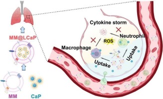
Neutrophils, which are highly motile, are the first arrivals of the body in response to inflammation [13]. As to the metastasis events, including premetastatic niche formation, seeding, intravascular neutrophil extracellular traps (NETs) and metastasis evolvement, neutrophils play an intricate and complex role [14]. However, neutrophils are found utmost difficulty engineered by genome editing due to the absence of CD68 promoter [15], while simply coating with neutrophil membrane onto nanoparticles would lose the inherent active chemotactic properties. In-situ surficial and temporary ornament onto living neutrophils are thus believed inherited with the metastasis-chemotaxis and promising in efficiently delivering cargos to inflamed tissues to overcome the intrinsic barriers (eg. plasma-peritoneal-barrier) that greatly restricts the exposure of diagnostic reagents from blood [16]. It has been well recognized that formyl peptide receptors (FPRs) were over-expressed on the activated neutrophils' surface to increase their sensitivity towards the stress signal [17]. All these characteristics suggest neutrophils might be able to assist the in vivo substrate transport by targeting FPRs to the sites with infection and inflammatory disorders.
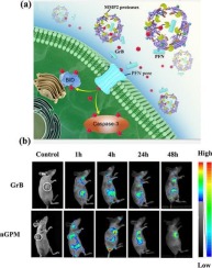
In this work, we develop a chemiluminescent probe which can specifically respond to O2− to trigger the photon release (See Scheme 1). The probe is “clicked” with a cFLFLF peptide (namely Peptide-CL) that can bind the FPR2 receptor overexpressed on neutrophils' membrane. Once injected, Peptide-CL can be effectively anchored onto the intrinsic circulated neutrophils. The in-situ modified neutrophils, inherited with the inflammation chemotaxis capability, could escort the silenced probe to the foci characterized with inflammation. Triggered by the local O2− at a high level, 510-nm photons will be spontaneously released after a self-immolation process. The neutrophil-assisting O2−-activatable chemiluminescent probe thus can specifically recognize inflamed tissues and provide reliable information for diagnosis.
留言 (0)