
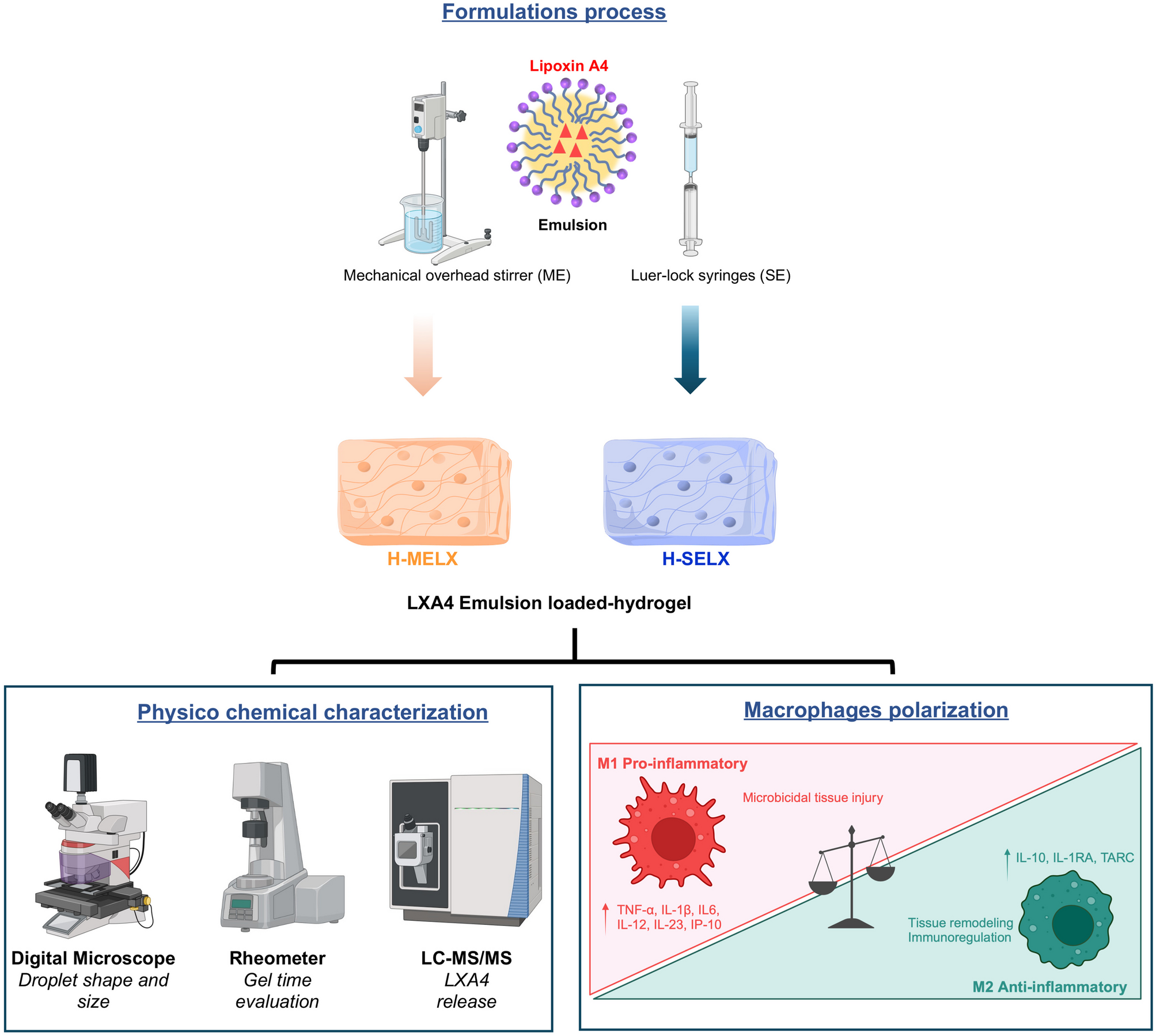
記住我

The colloidal properties of different ES100 nanoparticles are shown in Table 4. BUD-ES-NPs were prepared with a mean size of 144.7 ± 3.2 nm. NPs showed a negative ZP (− 22.6 mV) ascribed to the characteristic dissolution and ionization of ES100 at ⁓pH 7.0 [34]. The high ZP provides an electric repulsion thus preventing particle aggregation. Positively charged NPs were prepared by adding CTAB to the formulation. A significant increase in PS (p ≤ 0.05) was observed with a shift to a positive ZP of 33.3 mV. The positive charge induced allowed for HA coating. This was confirmed by the increase in PS (239.5 ± 9.2 and 274.8 ± 2.9 nm for BUD-ES-NP + and HA-BUD-ES-NP, respectively) (Fig. 1a and b) and the negative charge observed following coating (− 24.6 ± 2.8 mV). For all formulations, the PDI was inferior to 0.3 which demonstrated formulation monodispersity.
Table 4 Physicochemical properties of ES100 nanoparticles (n = 3)Fig. 1
Particle size distribution by intensity curve of a BUD-ES-NP + and b HA-BUD-ES-NP and SEM images of c BUD-ES-NP + and d HA-BUD-ES-NP. Magnification × 15 K, scale bar represents 1 µm
Efficient encapsulation of BUD was attained with %EE exceeding 98% (Table 1) and an average drug payload of ⁓17.6%. The high %EE is attributed to the lipophilic nature of BUD (Log p value 1.9) and hence low affinity to water [35].
Morphological examination of BUD-ES-NP + and HA-BUD-ES-NP using SEM (Fig. 1c and d, respectively) showed smooth spherical nanoparticles. The size was uniform with no aggregates. HA-BUD-ES-NPs have slightly higher PS compared to the uncoated formulation. These results are comparable to those determined by DLS.
In vitro drug releaseThe pH-dependent release profile of BUD from ES100 nanoparticles under sink conditions was studied and compared to BUD suspension (Fig. 2). At pH 1.2, BUD showed a high rate of drug release; 70% during the first hour and exceeded 90% at 2-h intervals. On the other hand, BUD release from BUD-ES-NP + was highly sustained with a release of ⁓29% at 1 h probably corresponding to the surface-entrapped drug and ⁓30% after 2 h. At pH 6.8, a spurt in drug release was observed (⁓60% at 4 h). This reflects the pH-responsive dissolution of the polymer allowing for drug release at the site of inflammation. HA coating of ES-NPs resulted in a statistically insignificant (p > 0.5) reduction in BUD release rate compared with BUD-ES-NP + .
Fig. 2
Release profile of budesonide from BUD-ES-NP + and HA-BUD-ES-NP formulations in pH 1.2 and 6.8 at 37 °C over 24 h (n = 3)
Storage stability of ES100 nanoparticlesBUD-ES-NP + and HA-BUD-ES-NP formulations were stored at 4 °C. At 2-week intervals, PS and ZP were measured and compared to zero time (Fig. 3). Both formulations showed a slight progressive increase in size over time. In the case of BUD-ES-NP + , the increase was significant (p ≤ 0.05) reaching a maximum of 25% after 8 weeks. On the other hand, the HA-coated formulation increase in size was statistically insignificant (p > 0.05) with a maximum increase of 18%. Regarding PDI, both BUD-ES-NP + and HA-BUD-ES-NP showed an increase in PDI which remained below 0.4 indicating that the formulations relatively retained their mono-dispersity upon storage. Whereas storage affected PS, no change in ZP was observed for both formulations. Stable ZP, especially for the HA-coated formulation, reflects the stability of the coat over time which is important for targeting inflammation.
Fig. 3
Change in particle size (nm) and zeta potential (mV) of BUD-ES-NP + and HA-BUD-ES-NP upon storage at 4 °C over 8 weeks (n = 3)
Stability in simulated gastrointestinal fluidsIn view of the planned oral administration of ES100 nanoparticle formulations, in vitro stability in simulated gastrointestinal fluids was performed (Table 5). In SGF, both formulations showed a significant increase in size (p ≤ 0.05). The increase was more pronounced for the HA-coated formulation (21% and 70% increase in PS for BUD-ES-NP + and HA-BUD-ES-NP after 2 h incubation, respectively). Also, a change in ZP was observed following incubation in SGF (28% and 40% decrease in ZP for BUD-ES-NP + and HA-BUD-ES-NP after 2 h incubation, respectively). A reverse pattern was observed in SIF where the change in PS and ZP was higher for BUD-ES-NP + than HA-BUD-ES-NP (109% and 37% increase in size with 56% and 49% decrease in ZP for BUD-ES-NP + and HA-BUD-ES-NP, respectively). The change in colloidal properties of ES-NPs could be explained by the formation of protein corona around the particles which is accompanied by alteration in size and surface charge [36]. This interaction is mainly driven by hydrogen bonds and van der Waals forces as previously described by Wang et al. [36]. The composition of protein corona is influenced by particle size, shape, and surface properties such as zeta potential, hydrophobicity, and functional groups [37] thus explaining the difference in behavior observed between BUD-ES-NP + and HA-BUD-ES-NP. One of the main aims of the experiment was to investigate the stability of the HA coat in gastrointestinal fluids. Retention of negative surface charge following incubation in simulated gastrointestinal fluids reflects the stability of the HA coat on the surface of positively charged BUD-ES-NP + .
Table 5 Stability of ES100 nanoparticles in simulated gastric (SGF) and simulated intestinal (SIF) fluids in terms of change in particle size (PS) and zeta potential (ZP) (n = 3)In vitro cell culture studiesCaco-2 cell viability assayCaco-2 cells were exposed to blank formulations and to ES100 formulations standardized at increasing BUD concentrations. Cell viability was calculated relative to control (Fig. 4). The drug solution showed dose-dependent toxicity on Caco-2 cells. As the concentration of the drug solution increased from 0.1 to 3.2 µg/ml, the cell viability decreased from 99 to 30%. On the other hand, in the drug concentration range tested (0.1–3.2 µg/ml), BUD-ES-NP + and HA-BUD-ES-NP showed cell viability exceeding 90% as obtained for blank formulation. The results reflect that loading of BUD into ES-NPs reduced drug cellular toxicity and enhanced safety on intestinal cells.
Fig. 4
Percent Caco-2 cell viability after exposure to BUD-ES-NP + and HA-BUD-ES-NP compared to blank formulations and BUD solution. Data expressed as mean ± SD (n = 3)
Cellular uptakeConfocal laser microscopy scan was used to study cellular uptake of C6-loaded formulations in Caco-2 cells compared to free dye solution (Fig. 5A). Green fluorescence corresponds to C6 whereas the cell nuclei marker, DAPI, is observed as blue fluorescence signals. A quantitative assay of cellular uptake was done by calculating cellular fluorescence intensity via ImageJ software (Fig. 5B). C6 solution showed sparse fluorescence signals. Reduced Caco-2 uptake of C6-free solution has been previously reported and attributed to the inability of raw C6 to be directly internalized by the cells [38]. Loading of C6 into ES-NPs resulted in significantly higher fluorescence intensity (p ≤ 0.05). Moreover, the HA coating of the nanoparticles showed a further increase in fluorescence intensity in the cytoplasmic and nuclear areas. The results support the assumption that nanoparticle physicochemical properties and surface nature in contact with cells or cellular components affect cellular uptake [38, 39]. This is ascribed to the difference in cell penetration mechanisms [40]. Hence, surface modification of nanoparticles could successfully provide enhanced cellular interaction [41]. C6-ES-NP + are mostly internalized by endocytosis and passive targeting [40]. Also, the positive charge on their surface could benefit the interaction with the negatively charged cell membrane [42]. HA coating of the nanoparticles significantly improved cellular uptake which is mostly due to HA interaction with its major cell surface receptor CD44, consequently, resulting in nanoparticle internalization via receptor-mediated endocytosis. The specific affinity of HA to CD44 makes it an ideal targeting moiety for increasing cellular uptake and concentration of drugs at the surface of cancerous or inflamed colonic cells overexpressing this receptor [40, 43].
Fig. 5
Cellular uptake of C6-ES-NP + , HA-C6-ES-NP, and coumarin-6 solution in Caco-2 cells: A confocal laser scan microscope images. Blue and green fluorescence signals represent the cell nuclei (DAPI) and coumarin-6, respectively, and B fluorescence intensity is calculated using ImageJ software. Data expressed as mean ± SD. ap ≤ 0.05 vs coumarin-6, bp ≤ 0.05 vs C6-ES-NP + , and cp ≤ 0.05 vs HA-C6-ES-NP
Inflammatory markers expressionLipopolysaccharide (LPS) is considered one of the most potent inducers of gut inflammation [44]. It was shown that LPS can initiate a cascade of signal transduction through Toll-like receptor 4 extracellular domain binding, thus enhancing pro-inflammatory cytokine production [45] and hence is commonly used to induce cellular inflammation. Following induction of inflammation in Caco-2 cells, IL-8 and TNF-α production in the culture medium were determined using ELISA kits. As shown in Fig. 6, LPS resulted in a marked increase in IL-8 and TNF-α levels (12 and 20-fold increase, respectively, compared to LPS untreated cells). BUD treatment resulted in a significant decrease in cytokines levels (25% and 28% decrease in IL-8 and TNF-α, respectively, p ≤ 0.05) as it exerts a direct anti-inflammatory effect on intestinal epithelial cells [46]. Loading of BUD into ES-NPs + resulted in a further decrease in IL-8 and TNF-α levels (46% and 50%, respectively, p ≤ 0.05). Maximum reversal of LPS inflammatory effect was observed following treatment with HA-BUD-ES-NP (63% decrease in both IL-8 and TNF-α compared to LPS-treated cells). This could be explained by the enhanced cellular uptake achieved by the HA-coated NPs. Similarly, HA-functionalized polymeric nanoparticles [47] and BUD-loaded self-assembled HA-NPs [48] demonstrated higher anti-inflammatory effect as was shown by the reduced IL-8 and TNF-α secretion in inflamed cell models.
Fig. 6
Evaluation of inflammatory markers; A interleukin-1ß (IL-1ß) and B tumor necrosis factor-alpha (TNF-α) following inflammation induction in Caco-2 cells using LPS and treatment with BUD, BUD-ES-NP + , and HA-BUD-ES-NP compared to suitable controls. Values were expressed as mean ± SD (n = 5). ap ≤ 0.05 vs control, bp ≤ 0.05 vs LPS, cp ≤ 0.05 vs BUD, dp ≤ 0.05 vs ES-NP + , ep ≤ 0.05 vs HA-ES-NP, fp ≤ 0.05 vs BUD-ES-NP + , and gp ≤ 0.05 vs HA-BUD-ES-NP
In vivo efficacyAcetic acid-induced colitis shares several clinical features of human IBD. It mimics human pathophysiology in cytokine profile and histopathological attributes and is characterized by infiltration of neutrophils and subsequent colon tissue damage via reactive species formation [49, 50]. Also, its simplicity, reproducibility, and the rapid appearance of inflammation and clinical parameters make it a popular experimental model for IBD [51].
Effect on colon length, DAI, and macroscopic ulcer scoreIBD induction by acetic acid led to a significant decrease in colon length (10 ± 1.5 cm) associated with a significant increase in DAI (5.4 ± 0.5) and macroscopic ulcer score (4.6 ± 0.5) in the positive control group versus the negative control (14.8 ± 0.2 cm, 0, 0, respectively; p ≤ 0.001 for the three parameters) (Fig. 7A–D). All these changes were significantly improved following treatment with either BUD, BUD-ES-NP + , or HA-BUD-ES-NP versus the positive control with p ≤ 0.001 regarding both DAI and ulcer score when comparing the three treated groups to the positive control one. However, when comparing the colon length in BUD, BUD-ES-NP + , and HA-BUD-ES-NP-treated groups versus the positive control one, p values were ≤ 0.05, ≤ 0.01, and ≤ 0.001, respectively. The highly significant changes observed indicated mucosal healing and alleviation of inflammation [52]. It is worth mentioning that HA-BUD-ES-NP presented the best recovery with p ˃ 0.05 regarding colon length and ulcer score when compared to the negative control group.
Fig. 7
A Macroscopic evaluation of colitis, B colon length, cm, C disease activity index (DAI), and D ulcer score following treatment of acetic acid-induced colitis with BUD, BUD-ES-NP + , and HA-BUD-ES-NP compared to suitable controls. Values were expressed as mean ± SD (n = 5). ap ≤ 0.05 vs negative control, bp ≤ 0.05 vs positive control, cp ≤ 0.05 vs BUD, dp ≤ 0.05 vs BUD-ES-NP + , and ep ≤ 0.05 vs HA-BUD-ES-NP
Histopathological changes assessment by H&E stainingHistopathological examination of H&E-stained colon tissue sections was done (Fig. 8A). In comparison to the normal histological appearance shown in the negative control group, the examination of positive control group sections revealed a complete loss of normal structure of colonic mucosa. Sections showed extensive necrotic destruction of the epithelium with hemorrhage, edema, crypt damage, ulceration, and vacuolation at mucosal and sub-mucosal layers. In addition, severe inflammatory cellular infiltration was also noted. The loss of normal colonic mucosal structure is still seen in sections from BUD-treated group. Areas of epithelial necrotic destruction, some scattered hemorrhage, edema, crypt damage, and small mucosal ulcers were still present. Moreover, cellular inflammatory infiltration was evident. On the other hand, BUD-ES-NP + and HA-BUD-ES-NP-treated groups showed substantial inflammation subsidence with the almost normal structure of colonic mucosa and mild inflammatory cellular infiltration. Small areas of ulceration, vacuolation, and edema were still seen. However, almost normal crypts and submucosa were seen in the HA-BUD-ES-NP-treated group.
Fig. 8
A Histopathological examination of H&E-stained colon tissue sections of negative control showing normal colonic mucosa. Regularly and parallelly arranged crypts (black arrows) perpendicular to the muscularis mucosae consisting of absorptive cells, goblet cells, and endocrine cells (red arrows). The lamina propria contains a variable number of inflammatory cells and a rich network of capillaries, venules, and lymphatics (blue arrows), and normal submucosa (green arrows). Positive control showed complete loss of normal structure of colonic mucosa with extensive necrotic destruction of the epithelium, hemorrhage, edema, crypt damage (red arrows), and ulceration and vacuolation at mucosal and sub-mucosal layers (green arrows), in addition to severe inflammatory cellular infiltration (black arrows). The BUD group showed loss of normal structure of colonic mucosa with necrotic destruction of the epithelium which is still seen. Also, some scattered hemorrhage, edema, crypt damage (red arrows), and small areas of ulceration and vacuolation at mucosal and sub-mucosal layers (green arrows), in addition to severe inflammatory cellular infiltration (black arrows). The BUD-ES-NP + group showed an almost normal structure of colonic mucosa with mild inflammatory cellular infiltration (black arrows) and a small area of ulceration and vacuolation (green arrows) and edema (red arrows) with almost normal crypts. HA-BUD-ES-NP showing normal colonic mucosa with a small area of ulceration and vacuolation (red arrows) and mild inflammatory cellular infiltration (black arrows) and normal submucosa (green arrows). The scale bar represents 50 µm. B The histopathological score of treated groups following administration of BUD, BUD-ES-NP + , and HA-BUD-ES-NP compared to control. Values were expressed as mean ± SD (n = 5). ap ≤ 0.05 vs negative control, bp ≤ 0.05 vs positive control, cp ≤ 0.05 vs BUD, dp ≤ 0.05 vs BUD-ES-NP + , and ep ≤ 0.05 vs HA-BUD-ES-NP
Based on the above findings, a histopathological score was calculated (Fig. 8B). This was significantly increased in the positive control group (4.83 ± 0.29) versus the negative control one (p ≤ 0.001). Yet, it was significantly improved following treatment with either BUD (4.26 ± 0.16), BUD-ES-NP + (3 ± 0.29), or HA-BUD-ES-NP (2.26 ± 0.47) with p ≤ 0.001 for both BUD-ES-NP + and HA-BUD-ES-NP-treated groups versus the positive control.
Expression of inflammatory markers in colonic tissue homogenateSince inflammation plays a major role in the pathogenesis of acetic acid-induced colitis, the expression of inflammatory markers in colonic tissue homogenate was assessed. The selection of inflammatory biomarkers was based on previous studies, where TNF-α expression was found to accelerate the inflammatory cascade through nuclear factor (NF-κB) pathway activation and hence is directly involved in colon tissue destruction [53, 54]. Also, McAlindon et al. demonstrated the evident role of IL-1β in IBD [55]. The role of the selected inflammatory biomarkers was confirmed by the significant increase in IL-1β and TNF-α (p ≤ 0.05) in the positive control group versus the negative control one (2.6 and 4.8 folds increase, respectively) (Fig. 9A). This significant elevation further explains the marked inflammatory cellular infiltration observed in the histopathological sections of the positive control group (Fig. 8A). Treatment with BUD, BUD-ES-NP + , and HA-BUD-ES-NP led to 37%, 56%, and 65% decrease in IL-1β respectively when compared to the positive control group (Fig. 9A). A similar pattern was observed for TNF-α expression which also showed a significant decrease of 38%, 52%, and 69% following treatment with BUD, BUD-ES-NP + , and HA-BUD-ES-NP, respectively, compared to the positive control group (Fig. 9B).
Fig. 9
Evaluation of various biomarkers in colonic tissue; A interleukin-1ß (IL-1ß, B tumor necrosis factor-alpha (TNF-α), C colonic E-cadherin (E-cad), and D colonic miR-21 expression in acetic acid-induced colitis following administration of BUD, BUD-ES-NP + , and HA-BUD-ES-NP compared to control. Values were expressed as mean ± SD (n = 5). ap ≤ 0.05 vs negative control, bp ≤ 0.05 vs positive control, cp ≤ 0.05 vs BUD, dp ≤ 0.05 vs BUD-ES-NP + , and ep ≤ 0.05 vs HA-BUD-ES-NP
Expression of colonic E-cadherinThere is a clear link between IBD etiology and deficits in gastrointestinal epithelial barrier function. An intact gut barrier protects against leakage of antigens from the intestinal lumen into the interstitial space and the lamina propria. It also controls polymorphonuclear leukocytes (PMN) migration across the epithelium into the lumen. A pivotal component of the epithelial adherens junction, E-cadherin, was shown to play a crucial role in cell–cell adhesions which are fundamental to intestinal epithelial barrier function [56]. Previous studies demonstrated mutations and changes in the E-cadherin gene in UC patients [57, 58]. Also, a dominant interfering E-cadherin mutant in the intestinal epithelium was shown in IBD-affected mice [59]. Moreover, the functional involvement of cytoplasmic E-cadherin-associated protein p120-catenin in IBD is well reported [60, 61]. In the current study, IBD induction by acetic acid resulted in a 45% decrease in E-cadherin in the positive control group versus the normal negative control one with p ≤ 0.001 (Fig. 9C). However, following treatment with BUD, BUD-ES-NP + , and HA-BUD-ES-NP, a respective increase of 16%, 41%, and 70% in E-cadherin level was observed compared to the positive control group (p ≤ 0.05 for BUD-treated group, p ≤ 0.001 for BUD-ES-NP + and HA-BUD-ES-NP-treated groups). Moreover, restoration of E-cadherin expression was achieved by HA-BUD-ES-NP showing an insignificant difference compared to its level in the negative control group (p > 0.05).
Colonic miR-21 expressionOne of the most widely studied miRNAs regarding health and disease is miR-21. Regarding IBD, miR-21 level elevation is suggested to be a pathological finding [62, 63]. Our results showed that IBD induction by acetic acid led to a significant 2.4 folds increase in colonic miR-21 expression in the positive control group versus the normal negative control one with p ≤ 0.001 (Fig. 9D). This is in accordance with other studies showing that miR-21 ablation in mice is protective against DSS-induced colitis [64, 65]. Also, miR-21 downregulation was reported in UC patients in the remission phase [66]. However, treatment with BUD, BUD-ES-NP + , and HA-BUD-ES-NP led to 19%, 44%, and 63% significant decrease in colonic miRNA-21 expression in the three treated groups, respectively, when compared with the positive control group (p ≤ 0.001 for the three groups). Furthermore, miRNA-21 expression in HA-BUD-ES-NP-treated group was insignificantly different from its level in the negative control group with p > 0.05.
The role of miR-21 in IBD pathogenesis could be attributed to its effect on intestinal inflammation. Herein, we found a strong positive correlation (R2 > 0.9) between miR-21 expression and inflammatory markers, TNF-α and IL-1β (Fig. 10A and B). In accordance with our results, the miR-21 expression on immune cells with the promotion of inflammatory cytokines production has been previously reported [67, 68]. Another possible effect of miR-21 in IBD pathogenesis is its effect on gut permeability where a negative correlation between E-cadherin and miR-21 levels was previously reported [69, 70]. Similarly, a strong negative correlation between colonic miR-21 expression and colonic E-cadherin level with a determination coefficient of 0.98 was observed in the current study (Fig. 10C). Recently, one study showed exacerbation of dextran sulfate sodium (DSS)-induced colitis in mice due to the uptake of exosome-derived miR-21a-5p from abnormally polarized macrophages by intestinal epithelial cells and a decline in E-cadherin level [71].
Fig. 10
Correlation between levels of A and B inflammatory markers (TNF-α and IL-1ß) or C E-cadherin (E-cad) and miR-21 expression
The combined results of the in vivo tests performed on the acetic acid-induced IBD animal model all showed the enhanced efficacy of BUD upon loading into NPs. Indeed, the small size of nanocarriers was previously shown to allow for more efficient drug targeting to affected tissues through the eEPR effect which allows for accretion at the inflamed and disrupted epithelium [72]. Also, inflamed tissues show epithelial barrier disruption, in addition, to an increase in the production of mucus and immune cell infiltration [
留言 (0)