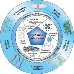
Hydrogels are 3D cross-linked porous hydrophilic polymer scaffolds with exceptional characteristics such as swelling ability, biocompatibility, self-healing, self-adhesiveness, and stimuli sensitivity [1]. By definition, water must make up 10–20% of the product's dry weight to be called a hydrogel. Hydrogel's 3D-network structures can be formed by covalent crosslinking, hydrogen bonding, ionic forces, biorecognition interactions, hydrophobic interactions, physical entanglement of polymer chains, or a combination of two or more of the above interactions [2]. Since the discovery of hydrogels by Wichterle and Lím in 1960 [3], a wide range of biomedical applications including drug delivery, wound healing, tissue engineering, implantable devices, and wearable biosensors have been discovered [4]. To date, natural polymers, conductive synthetic polymers, crosslinkers, fluorescent agents, and buffer solutions, as well as nanomaterials (carbon, metal, and metal oxide NPs), have been utilized to produce hydrogels with specific stimuli for sensing applications [5]. Therefore, new types of hydrogels such as injectable, fluorescent, electrically conductive, and stimuli-responsive, self-healing, shape-memory hydrogels have been developed to meet clinical needs [6].
Over the last few decades, medical diagnoses have been performed in biofluids (e.g., urine, serum, plasma) and organ tissues containing high concentrations of the ions, biomolecules, and biomarkers of interest [7,8]. However, the increasing demand for noninvasive diagnostic devices based on other biofluids such as sweat, tears, and saliva, which contain much lower concentrations of biomarkers than the bloodstream, poses a greater challenge for the development of more sensitive and selective sensing platforms [9]. Various protocols for immobilizing hydrogels with specific bioreceptors (e.g., antibodies, aptamers, and enzymes) have been developed and optimized, as wearable and implantable scaffolds. The bioreceptors sometimes act as cross-links and form dense network architectures, while the analytes react with these bioreceptors and cause physical changes (swelling) [10]. Redox-responsive hydrogels that promote rapid electron transfer have been extensively studied for the electrochemical detection of inflammatory biomarkers released from the skin [11]. Reactive hydrogels offer unique features, such as tunable naked-eye detection, real-time analysis, and the ability to function without a power supply. However, the main challenge is large-scale fabrication and optimization for the development of a sensor for a particular analyte [12].
Advances in wearable and implantable biosensors would benefit from the use of hydrogels because they are biocompatible, self-adhesive, easy to process, have strong sensing capabilities and rapid responsiveness (swelling, electrical conductivity, and fluorescence), and can extract biomarkers in real time from skin and tissue [4,13]. These hydrogels can simulate the mechanical, physicochemical, and biological properties of innate tissues. Compared to other biomaterials, hydrogels can resemble living tissues and are inherently similar to the components of the extracellular matrix. Interestingly, they provide a suitable environment for cell survival and growth and can reduce the mechanical effects on tissues after water absorption [14]. Hydrogels enable the development of wearable tattoo sensors that interact with human skin or organs to provide sensitive and rapid detection of human metabolites [6]. Hydrogels also facilitate the extraction of biomarkers in biofluids (wound exudate) produced by various pathological skin conditions [15]. Tuning the functionality of hydrogels can also increase the loading capacity and accessibility of target biomarkers (e.g., ions, drugs, bacteria, viruses) to bioreceptors immobilized on hydrogels, resulting in a lower background signal due to less nonspecific binding with other interfering molecules [16].
This review covers the basic concept of hydrogel-based biosensors, immobilization of bio-recognition elements (e.g., antibodies, enzymes, and aptamers), and extraction of biomarkers (ions, drugs, proteins, enzymes, hormones) from skin and biofluids, and hydrogel response. Detection of small biomolecules such as glucose, urea, lactate, and cholesterol, as well as pathogen detection, wound bioassay, tissue engineering, and cancer monitoring, are just some of the advanced uses of implantable, wearable, and disposable biosensors focused on in this article. In addition, the challenges of developing multi-stimuli-responsive hydrogels, the role of various nanomaterials, and their applications in biosensors are discussed. Market size, current challenges, and future research directions for hydrogels are proposed, offering new insights into the development of stimuli-responsive hydrogel-based biosensors.
In an aqueous medium, hydrogels are composed of a dense network of interconnected polymer chains that are hydrated. Other than water, a variety of solvents can be absorbed by hydrogels, and the functional groups present in the gel network (-COOH, -OH, -NH2, -CONH2, and -SO3H) control the gel's ability to absorb water or solvents [15]. The functionality of repeating polymer chains, monomers, and crosslinkers is responsible for the unique properties of hydrogels, and they can be chemically modified to adjust the hydrogel to acquire the desired properties. For example, natural polymers (e.g., cellulose, chitosan, gelatin, alginate, and albumin) exhibit pH-dependent behavior and are widely used for drug delivery [19]. However, these polymers show weak mechanical strength and electrical conductivity, and many undesirable responses to environmental stimuli. Increasing polymer concentration is a way to increase the mechanical strength of natural polymers, but it typically results in poor biocompatibility and decreased degradability [20]. Several studies have attempted to improve natural polymer-based hydrogels by incorporating synthetic polymers and nanoparticles, lamellae, and buffers into the formulation [21]. However, synthetic polymers are less environmentally friendly due to the monomers, crosslinkers, initiators, solvents, and polymerization byproducts generated during their production, which are usually not biocompatible or degradable.
Stimuli-bioresponsive hydrogels are smart materials that can drastically change their shape, volume (swelling, shrinkage, solvent receptivity), and other properties (light emission, light absorption, electrochemical behavior) in response to biological stimuli (such as glucose, ATP, enzymes, antibodies, etc.) [22]–[24]. Stimuli-bioresponsive hydrogels can be divided into physically crosslinked and chemically crosslinked hydrogels depending on the manufacturing process (See Fig. 1):
(1)
Physically crosslinked hydrogels are also referred to as “reversible hydrogels” because they can dissolve when environmental variables such as pH, ionic strength, and temperature are changed [25]. Temporary crosslinks are formed by the entanglement of polymer chains or by physically induced gelation caused by stimuli such as ion-ion interactions, hydrogen bonding, thermally driven gelation, complementary bonds, inclusion complexes, or hydrophobic interactions. Physically crosslinked hydrogels are less stable than chemically crosslinked hydrogels due to their reversible physical interactions and resulting mechanical properties [26]. They function inconsistently in vivo because they cannot be modified by gelation time, gel pore size, chemical functionalization, and degradation or dissolution. In addition, physically cross-linked hydrogels can enhance non-specific binding in sensory systems.
(2)
Chemically crosslinked hydrogels are also referred to as “permanent hydrogels” because their basic building blocks are crosslinked by covalent bonds. Compared to physically cross-linked hydrogels, chemically crosslinked hydrogels have areas of high cross-link density and are therefore very stable against degradation [27]. Thus, introducing a certain degree of chemical bonding into physically crosslinked hydrogels could be a useful technique to improve their strength.
Functionalization of bio-responsive hydrogels enables real-time response to various external stimuli and label-free detection compatible with readout devices. Their structures, compositions, biological functions, biodegradability, mechanical stability, viscoelasticity, and pH stability can be achieved and controlled by the spatial functionalization of hydrogels with specific biological entities to produce the desired stimuli. They are suitable as sensing materials for naked-eye detection because the color-providing substance is integrated into the hydrogel. For example, in the in vitro detection of glucose [28], a hydrogel was used to include the Shinkai receptor and Au-NP as optical indicators. Low glucose concentrations cannot be detected by the conventional assay, but by including Au NPs in the hydrogel, it is possible to convert low glucose concentrations into a visible signal (see Fig. 2).
However, the main challenge is large-scale fabrication and optimization for multi-analyte detection [12]. For example, Sikes et al. developed a polymerization-based amplification to detect influenza antigens [29]. In the presence of influenza A, a solid cross-linked polymer formed, and the formation of this solid polymer was indicative of the binding of the influenza A nucleoprotein. This study is critical because the antigen (influenza A) can be detected without any instrumentation. The swollen properties of hydrogels can also be used as biosensors [30]. In addition to the physical and mechanical changes, the analytes (fluorescent dyes or quantum dots) can be quantified. The targeted analyte (antigen) can also be detected. Antigen/antibody conjugated with fluorescent molecules in a hydrogel can lead to a shift in the wavelength and intensity of fluorescence, and these changes are proportional to the concentration of the analyte [23,24].
A molecular species, such as an enzyme, gene, antibody, nucleic acid, or biological system, such as a cell or organ, that uses a biochemical mechanism to interact with the analyte is referred to as a “bioreceptor.” [31]. In proportion to the interaction between the bioreceptor and the biomarker, a transducer generates a quantifiable signal. Various immobilization techniques have been described for coupling the bioreceptors to hydrogels. However, the selection of an appropriate immobilization approach, availability of reactive groups, monitoring of bioreceptor degradation, or operational stability must all be considered. The best-known method for stabilizing bioreceptors is the use of polyelectrolytes to increase the stability of certain enzymes and antibodies [6]. Several studies have been proposed for the stabilization of bioreceptors and microorganisms in biosensors, including vacuum drying, lyophilization, and encapsulation. New methods need to be developed to monitor the porosity of the hydrogel, as biomolecules trapped in the large pores tend to become inactive or cause bioreceptor leakage during immobilization [32]. Further research could be conducted to prevent the destruction of the hydrogel during the detachment of the bioreceptor during irreversible immobilizations.
There are two ways to immobilize bioreceptors in a hydrogel: physical (reversible) and chemical (irreversible) immobilization. Physical (reversible) immobilization of bioreceptors (e.g., electrostatic bonding or repulsion, van der Waals forces, and hydrogen bonding) allows detachment of the bioreceptor but is very sensitive to changes in pH. This method is commonly used for the immobilization of enzymes and proteins [33]. Chemical (irreversible) immobilization prevents the bound bioreceptors from being released without affecting the microstructure of the hydrogel or the functionality of the bioreceptors [6]. This method prevents the bioreceptors from detaching from the polymer chain. Techniques for this type of immobilization include crosslinking, covalent bonding, and entrapment. However, the chemical immobilization methods have significant drawbacks, such as loss of bioactivity, which affects the sensitivity and selectivity of the biosensor. In addition, excessive crosslinking during biomolecule entrapment can interfere with analyte transport. Therefore, crosslinking must be carefully performed to prevent the leaching of the bioreceptors and allow the targeted biomarkers to reach and bind to the bioreceptors [34]. Fig. 3 shows the micro- and nanoscale structure of hydrogels and several strategies used to incorporate bioreceptors into hydrogels. Table 1 shows various examples of biomolecule immobilization methods and their detection methods.
Hydrogels are increasingly used as immobilization substrates for immunoassays. Compared to conventional sensing materials, non-rigid, porous, hydrated gels increase bioreceptor immobilization and target binding while reducing steric hindrance. Photopolymerization of monomer solutions in conjunction with antibodies or other large proteins can rapidly prepare antigen-specific hydrogels [35]. However, the major drawback of antibodies is their expensive and unstable production processes, which may ultimately affect the shelf-life of biosensors. Enzymes are an example of large proteins that can be used to detect or monitor various diseases such as diabetes, obesity, and cancer. Membranes or support matrices, electrostatic interactions, chemical crosslinking, and electrodeposition have been used in the past to immobilize enzyme molecules on or in hydrogels [36]. For example, Endo et al., have immobilized glucose oxidase (GOx) on the PAA hydrogel matrix using EDC-NHS coupling [37]. In another study by Imani et al., L-lactate was encapsulated by electrostatic interactions inside the chitosan [38]. Similarly, aptamer several approaches have been developed to increase the binding affinity of aptamers to hydrogels, including chemical modification, cooperative binding, and structural stabilization [39]. The binding affinity of the aptamers to the hydrogel as well as the targeted molecules have been modified by several approaches: (i) modification of nucleobases, (ii) replacement of backbone, and (iii) attachment of functional groups to achieve higher binding affinity.
Numerous hydrogel-based biosensors have been developed and commercialized for use in a variety of applications such as medical diagnosis and health monitoring. The interaction between the analyte and the bioreceptors is registered physicochemically and then converted into a detectable signal that is captured by the sensor platform. The hydrogels serve as substrates (coatings) or even act as a sensor or transducer platforms. To date, several strategies have been developed to produce a rapid stimuli-responsive hydrogel, including (1) modifying hydrogels with small biomolecules capable of forming specific bonds with antibodies, protein receptors, or other biomacromolecules to effect a macroscopic change in the hydrogel [40], (2) modification of hydrogels with enzyme-sensitive substrates, such as short peptides, leading to a chemical change in which bonds of the substrate molecule are made or broken, and (3) incorporation of biomacromolecules into the hydrogel structure that scavenges small biomolecules and convert them into molecules with different physical properties, such as acidity [41]. These changes can cause the hydrogel to inflate or collapse, and the signal can be used for biosensing [42,43]. Analytes can be detected using a biosensor based on electrochemical (e.g., current, potential, impedance) or optical (fluorescence) signals, depending on the operating principle of the sensor. The use of nanomaterial matrices as transducers for electrical or optical signals could increase the detection limit [42,43].


留言 (0)