
記住我

During this study, we sought to elucidate the role of DAH and/or sorafenib in ameliorating the anticancer efficacy of experimentally induced HCC by assessing the mechanism of action. In our study, DAH combined with SOR significantly enhanced its antiproliferative and cytotoxic effects on HepG2 cancer cells. HepG2 cells responded more positively to DAH and/or SOR since both compounds had lower IC50 values than WI-38 cells. This suggested that the combination of DAH and SOR was found to be more effective against HepG2 cells than either compound alone, suggesting that when used together, both compounds act synergistically in suppressing tumour growth. This further supports the notion that SOR is an effective treatment for hepatocellular carcinomas. After treating HepG2 cells with DAH at different doses, the IC50 values for SOR were significantly reduced. Our data showed that DAH showed the potential to act synergistically with SOR, thereby reducing the required dose. This could lead to a reduction of its side effects and improved efficacy, which would make SOR a safer and more effective chemotherapeutic drug for the potential treatment of liver cancer (Abdu et al. 2022).
Our data revealed that there was a significant increase in the levels of serum ALT, AST, and GGT in HCC mice, which could be explained by compromised cell membranes in certain types of hepatocytes. In line with current research findings, Huang et al. (2012), Badawy et al. (2021), and Abdu et al. (2022) have all reported similar findings. Accordingly, it was determined by Elzoghby et al. (2013) that both synthetic and natural anticancer medicines have the potential for antitumor activity when they have low liver damage enzyme levels. Furthermore, we found that treatment with DAH and/or SOR significantly reduced serum levels of these enzymes in HCC mice. The combination of DAH and/or SOR was the most effective treatment for reducing these enzymes. It is noteworthy that our results were in line with those of another study in which DAH was given to diabetic rats with type II diabetes to prevent hepatotoxicity (Heidari et al. 2022).
The primary gold standard for the detection of HCC is the AFP (Abdallah et al. 2022). In terms of AFP levels, the treatment with SOR and DAH was significantly associated with a decrease in levels, whether both treatments were administered alone or in combination with the highest level in HCC + SOR and HCC + DAH, respectively. Several studies have shown that the A. officinarum rhizome extract and its components are anticancer agents and that they possess potential anticancer activities against many cancer cell lines, such as the breast, neuroblastoma, liver, and lung (Basri et al. 2017).
In our study, it has been shown that DAH and/or SOR treatment decreased MDA and elevated T-SOD levels in HCC-bearing mice. The results of our research suggest that treatment with DAH and/or SOR significantly reduced oxidative stress levels. DAH, in accordance with this, stimulated T-SOD and decreased MDA levels in HepG2 cells, which supports this statement by enhancing free radical scavenging activity. As reported by Akazawa et al. (2006), the medicinal properties of this plant are mainly due to its DAH, which has diverse biological activities, including free radical scavenging, necessary for its therapeutic effects on cancer cells.
The docking results of our study demonstrated that DAH and SOR could bind to the active sites of CASP8 and MMP9 (Augoff et al. 2022), and the binding scores indicated that these compounds had a high affinity for the enzymes. Furthermore, the binding of the compounds to the promotor sequences of CASP8, IL-6, p53, MMP9, and VEGF suggested that the compounds could downregulate the expression of these genes. These findings were further supported by the biological analysis in our results, which exhibited that DAH and SOR could inhibit the activity of CASP8 and MMP9.
Apoptosis is triggered by numerous stressors like DNA damage and oxidative stress, as well as p53, which activates the death receptor cascade or mitochondria-mediated cell damage (Zhivotovsky and Orrenius 2010). This expression was significantly elevated after DAH and/or SOR therapy, either separately or together. The expression of the p53 gene may be triggered by the production of various DNA adducts, which too affect DNA replication, DNA helix distortion, and gene mutations. As a result of DAH treatment, HCC mice had higher p53 expression (Mandlik and Mandlik 2021).
The expression of caspases mediates apoptosis, which is caused by the cleavage of hundreds of different proteins by caspases (Lopez and a. TS, , 2015). The relevant data confirmed that CASP8 expressions were downregulated in the HCC group as compared to the control group. HCC + SOR + DAH had a dominant apoptotic impact compared to the HCC + SOR group, followed by the HCC + DAH group, and the HCC + DAH group had the lowest expression compared to the HCC untreated group. As a consequence of treatment with DAH and/or SOR, either in combination or individually, these expression levels significantly increased.
Some studies indicate that MMPs are a crucial component of regulating cell proliferation, invasion, and migration (Stetler-Stevenson and Anita 2001). In both cases, the expression of the MMP9 protein was significantly upregulated by treatment with DAH and SOR, either individually or in combination. Kim et al. (2006) reported that a DAH from the medicinal plant Alnus hirsuta dissuades the transcription of COX-2 and MMP9 when applied to cultured human mammary epithelial cells.
As a result of the treatment with either DAH or SOR alone or in combination, the expression of VEGF was significantly increased. Ryan et al. (1998) reported that VEGF is one of the most potent and definitive angiogenesis regulators that are crucial for solid tumour growth. According to Hu et al. (2019), a diarylheptanoid blockade VEGFR-2-mediated signalling cascades in human umbilical vein endothelial cells (HUVECs). This information may offer new insights into the possibility of a DAH as an anti-angiogenesis therapeutic agent.
IL-6 molecules are released from T cells and can induce B cell proliferation, differentiation, and antibody production (Liu et al. 2010). The expression of IL-6 was significantly downregulated following treatment with DAH and SOR either alone or in combination, with the highest expression in the HCC + SOR + DAH group followed by the HCC + SOR group and lowest expression in HCC + DAH group, as compared with the HCC untreated group. DAH may function as an anti-inflammatory drug by specifically inhibiting COX-2, the synthesis of cytokines involved in the signalling cascade of inflammation, or the modification of pro-inflammatory gene expression (Vanucci-Bacqué and Bedos-Belval 2021).
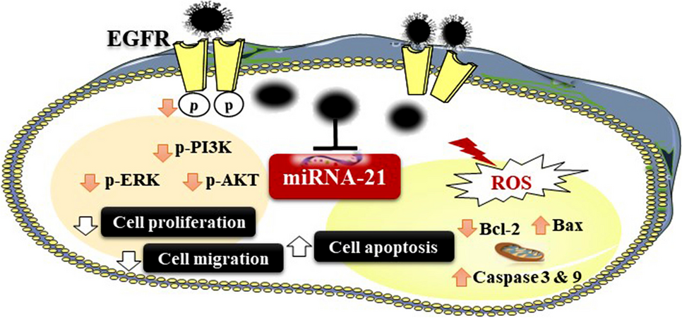
Therefore, these results supported our theory that DAH has an antitumor effect through targeting of p53 and MMP9 axis and that effect is critical to DAH-mediated HCC amelioration. Based on the study of HepG2 cells and in vivo studies, Fig. 9 summarizes the proposed antiproliferative and apoptotic action of DAH, as well as the downregulation of p53, IL-6, CASP8, MMP9, and VEGF genes in vivo by DAH as compared to control. The treated liver cancer cell line and hepatic genes were simulated to induce intrinsic and extrinsic pathways for apoptosis.
Fig. 9
A schematic diagram illustrates DAH’s potential mode of action in vivo. With DAH treatment, p53 and MMP9 levels were downregulated as well as GSH and T-SOD levels were increased in the DAB-induced HCC
Regarding the histopathological examination, our findings also revealed a focal area of necrosis surrounded by hepatocytes with acidophilic cytoplasm and hyperchromatic nuclei, and a congested portal vein, along with loss of hepatic architecture with the presence of a focal area of hepatocellular degeneration with pyknotic nuclei and acidophilic cytoplasm as well as vacuolar degeneration of hepatocytes surrounded by inflammatory cells in the livers of patients with HCC. By treatment with DAH and/or SOR, the liver structure of HCC-bearing mice dramatically improved, particularly among those that were also treated with DAH and SOR. These results are consistent with those of Pathak and Khuda-Bukhsh (2007) who demonstrated that DAH is capable of producing substances that can inflict cellular injuries on cells of the liver as well as other tissues.
留言 (0)