
記住我


Ten healthy individuals (five men and five women) were included in the study. All participants completed a questionnaire before entering the study (Table 1), including measurements to calculate body composition using the US Navy formula (Shaheen et al. 2019) and an estimation of their physical activity on a scale of 1–6 using the Grimby/Frändin activity scale stated as PAS (Grimby et al. 2018), 1 represents no physical activity and 6 intense work out several times a week. Participants aged between 18 and 75 years were eligible for inclusion. The exclusion criteria were pregnancy, skin ulcers, previous surgery on blood vessels of the lower limbs, pacemaker, intracardiac defibrillator, advanced heart disease, kidney failure, cancer and neuromuscular or metabolic disease. The study included a MP scan and sessions where Doppler ultrasound was performed during NMES stimulation to measure PVV, all while the participant was placed in a semirecumbent position.
Table 1 Characteristics of the participants (n = 10)M median, R Range, BMI body mass index, PAS Physical activity scale
Electrode-setupsIn this study, two different electrode setups for applying NMES to the calf were tested and compared regarding various outcomes. The electrode setups differed regarding electrode- type, and placement and were compared to investigate if textile electrodes knitted in fixed transverse positions in a sock, would yield a non-inferior increase of PVV from baseline compared to standard gel electrodes placed at individual MP.
The first electrode setup designated TTE, short for transverse textile electrode setup, consisted of two rectangular textile electrodes (2 × 2.5 cm) transversally knitted with the inner edges 1.8 cm apart into the back of a one-size-fits-all sock (polyamide/Lycra blended yarn) (Fig. 1a–b). The electrodes were placed approximately at the largest circumference of the calf with the electrodes positioned to cover areas of the calf, given the large interindividual variation of MP locations, which according to a MP map created by Botter et al. 2011 in general has a high likelihood of containing a MP (Botter et al. 2011). When the sock was applied to the calf the textile electrodes stretched to the size of approximately 3 × 3 cm, and the distance between the inner edges of the textile electrodes increased to approximately 3 cm. The same socks were used for all the subjects; therefore, the electrode size might alter slightly due to the stretch of the textile as well as the localization of the electrodes may change slightly. The electrodes were knitted into the sock using industrial intarsia knitting which allows for seamless integration of patterns of functional components in a single process. The material of the electrodes was silver coated polyamide multifilament yarn, with trade name Shieldex® (produced by Statex Produktions und Vertriebs GmbH). On the outside of the sock, the electrodes medial and lateral edges were covered with elastic yarn floats (polyamide/Lycra). The purpose of the floats was to hold a moisture-containing sponge (0.5 × 3 × 3 cm, injected with 2 ml NaCl 0.9 mg/ml) in direct contact with each of the underlying electrodes, to increase the local pressure and humidity of the electrode/skin-interface, and thus uphold an adequate electrical conduction (Fig. 2) (Euler et al. 2021a, b). Since the TTE-sock was only a prototype, a simple “off the shelf” melamine cleaning sponge was used as the moisture container. The different layers of the textile electrode are displayed in Fig. 2. Crocodile clips were used to connect the wires from the NMES device to the outside of the electrode fabric of the TTE.
Fig. 1
Displaying the transverse textile electrodes. Sock with knitted transverse textile electrodes, the face side (a–b), the back side with pink melamine sponges attached (c–d) and the sock displayed in an oblique view (b, d)
Fig. 2
The layers of the transverse textile electrodes. a A longitudinal view of an uncut textile electrode, and b a transversal view of a longitudinal cut through section displaying the layers of the textile electrode. For better visibility of the layers, some of the floats have been removed in Fig. 2a–b, as compared to Fig. 1c–d
The second electrode setup utilized commercially available standard gel electrodes (Compex Snap, Performance, DJO Global, USA, 5 × 5 cm) manually trimmed to squares sized 3 × 3 cm, to match the size of the TTE. Each standard gel electrode had a snap-on button to which each wire from the NMES-device was attached. The standard electrodes were placed on the skin areas of the calf, one on the medial side and one the lateral side, that required the least NMES current intensity to trigger a calf muscle response, i.e., the “best” MP, as determined by a standard motor point scan. This electrode setup was designated MPE, short for motor point electrode setup.
Motor point scanThe best MPs were found by scanning one half of the calf at a time (medial/lateral), using the NMES device’s 3 Hz sinusoidal wave motor scan program (Chattanooga Physio constant current generator, DJO Nordic, Malmoe, Sweden). Prior to the MP scan, the side of the calf about to be scanned was covered by a thin layer of conductive gel. A reference electrode (Compex Snap, Performance, DJO Global, USA, 5 × 5 cm) was placed on the contralateral side from the MP scan over the largest circumference of the calf, on a distance from the calf’s midline corresponding to 15% of the calf’s largest circumference. The definition of a MP was the same as in our previous study (Schriwer et al. 2023) e.g., as a location on the skin that resulted in a muscle twitch at the lowest level of stimulation compared to the closest surrounding area (Moon et al. 2012), and were determined by visual inspection and palpation of the muscle (Botter et al. 2011). Starting at the lowest NMES-level that was possible to set on the NMES-device (corresponding to 4 mA), the MP scan pen was used to search through one whole side of the posterior calf in accordance with the NMES-device instruction, during which time the examiner visually checked for any sign of a muscle twitch of the calf. If a visual muscle twitch was detected, the MP pen was held still in the location inducing the muscle twitch, and the muscle twitch was via manual palpation either confirmed as true or false. If true, the location on the skin underlying the tip of the MP scan pen was confirmed as a MP point (Gobbo et al. 2014). If no MP was found, the current intensity was increased by one NMES-level followed by a subsequent re-scan. This procedure was then repeated until a visible muscle twitch was detected in the calf, indicating the location of a MP, which was subsequently marked out on the scanned side of the calf (medial or lateral). All MP scans were performed by the same person to ensure that there was no examiner-bias.
NMES-SettingsFor both electrode setups, NMES was applied using the same NMES device as for the MP scan. The NMES stimulation used a biphasic symmetric square wave, meaning that the electrodes continuously were switching polarity so that they alternately, and for equally long durations, served as either anode or cathode. Thus, there was no designated anode or cathode in the electrode setups for this study. Based on previous studies on NMES discomfort, stimulation settings where set to 36 Hz frequency, 200 µs phase duration (400 µs pulse duration), 0.5 s ramp up time and 0.25 s ramp down time (Baker et al. 1993). The duration of each stimulation between the ramp up and ramp down time, i.e., the plateau time, was varied for different tests between 0.5 s, 1.5 s, 3 s and 5 s. The muscle rest between each cycle of combined ramp up, plateau time and ramp down, i.e., the OFF-time, was 8 s. The order in which the plateau times were tested was randomized. The NMES-level (0–999), representing a non-linear relationship to the current intensity ranging from 0 to 120 milliampere (mA), was gradually increased one NMES-level at a time as described below.
NMES Measurement Level I & IIThe NMES-device display the current used for the selected stimulation in NMES-levels ranging from 0 to 999, which in a non-linear pulse duration dependent fashion correlate to current amplitudes ranging from 0 to 120 mA. The formula to calculate this correlation may be obtained from the manufacturer (DJO Nordic, Malmoe, Sweden) upon reasonable request, but may not be publicly distributed.
When testing the two electrode setups, outcomes were registered at two distinct current intensities, designated measurement level I (ML I) and measurement level II (ML II). For every test performed, the current intensity was slowly increased one NMES-level at a time until a visible plantar flexion was induced. The current intensity needed to induce this plantar flexion was defined as ML I. A clearly visible plantar flexion was chosen as a point for outcome measure because, (1) it can be dichotomised; either you can see a plantar flexion, or you cannot, and (2) it will likely increase PVV compared to the baseline resting state (Clarke et al. 2006; Laverick et al. 1990). To avoid any examiner bias, only one examiner was used for all participants for the assessment of when a visible plantar flexion was induced by the NMES. ML II was defined as the current intensity corresponding to ML I plus an additional six NMES-levels increase on the NMES-device. For example, if plantar flexion was induced at NMES-level 20 (ML I), ML II would correspond to NMES-level 26 (= 20 + 6). The six-level NMES increase was chosen based on pre-testing indicating a significant increase of PVV compared to ML I, while any further intensity increases indicated to result in significantly more aborted test due to discomfort.
Since ML I and ML II could occur at different NMES-levels for different participants, correlating non-linearly to different mA amplitudes for different participants, we deemed the presentation of the testing procedure in mA amplitudes to be too unpractical to be easily reproducible in a clinical setting, since it would require the user to obtain the conversion formula and convert the mA amplitudes back into NMES-levels. Thus, only the NMES-levels are used to describe the different current intensities during the testing procedure, while the current intensities in the results are presented in mA for better relative comparisons in the statistical analysis. The range of increase in current between ML I and ML II is disclosed in the results section.
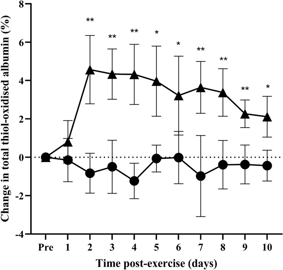
Hemodynamic measurementsUsing a Philips CX50 (2013) Doppler ultrasound machine (Philips Medical Systems, Andover, MA, USA), the widest accessible part of the popliteal and femoral veins was located and visualized in a longitudinal plane before beginning the hemodynamic measurements. The diameters of the veins were calculated at baseline. The diameter of the popliteal vein was 0.90 ± 0.29 cm and of the femoral vein 1.05 ± 0.26 cm. For the two electrode setups (TTE & MPE), PVV was measured in the popliteal and femoral veins at baseline (i.e., electrodes attached but no NMES administered), at ML I, and at ML II. For each subject in the study, all hemodynamic measurements were performed by the same ultrasonographist. During three consecutive NMES-stimulation cycles venous measurements were recorded, and subsequently the peak venous velocity (PVV) was assessed. The measuring tool on the ultrasound machine provided, when using doppler, the ability to save the recordings of blood flow in cm/s and measure the peak venous velocity after the stimulation was complete. The diameter of the vein during the stimulation was not calculated. PVV measurements during three NMES-stimulation cycles were performed and the mean of the three was used for statistical analysis. After analyzing the refill time of the veins, an eight second OFF-time was decided to be used between ON-times for the veins to be adequately refilled with blood before the next upcoming muscle contraction and PVV measurement. To quantify the potential benefit of NMES versus the baseline resting state, for each subject and setting, the percentual increase in PVV at ML I and ML II, as compared to PVV at baseline, was calculated and presented along with the absolute values. The formula used to calculate the percentual increase was:
$$\left( } \right) \, = \, \left( } \right) \left( } \right)} \right) / \left( } \right)} \right) \times 100$$
DiscomfortFor each stepwise increase in NMES-levels when testing the two electrode setups, participants were asked to fill in a form to rate their discomfort on a numerical rating scale (NRS) 0–10, where 0 was described to the subject as no discomfort and 10 as the worst imaginable discomfort (Hawker et al. 2011).
Statistical analysisThe sample size was determined prior to the start of the experiment based on a pilot study with a difference in PPV between ML I to ML II of 20 cm/s and sigma of 20, with the significance level set at p < 0.05 and power at 80% regarding the primary outcome, PVV in the popliteal vein. Based on the calculations, eight participants were needed to find a significant increase in PVV with an increase of the current intensity from ML I to ML II. We set the final sample size to ten participants.
The data were analyzed using SPSS version 27 (IBM Corp. Released 2016. IBM SPSS Statistics for Windows, Armonk, NY: IBM Corp.) in cooperation with a statistician. The four tested plateau times, 0.5, 1.5, 3 and 5. seconds, did not demonstrate any statistically significant difference regarding PVV or NRS, regardless of the electrode setup or measurement level. For this reason, the outcome values for PVV and NRS used in the final statistical analysis were based on all values, for all participants, obtained during the four different plateau times. Based on the relatively small number of participants and the Shapiro–Wilk indicating non normal distribution, the non-parametric Wilcoxon signed rank test was chosen to determine if there were any statistically significant differences between medians regarding current intensity (mA), PVV or NRS, for the two electrode setups. The data included some outliers, which were handled both using a rank-based statistics test and by adjusting the values of the outliers to the lowest/highest value within 1.5 times the interquartile range from the first respectively third quartile for the inferential statistics (Altman 1990). Analysis of correlations between subject characteristics and collected data were performed using Spearman’s ρ. Data are presented with median, interquartile range (IQR) and when comparisons were made the significance level for all analyses was set to p < 0.05. For the remainder of the text, “statistically significant” will be shortened as “significant”.
留言 (0)