
記住我


One hundred and twenty-four physicians responded to the survey. Most of the respondents were senior physicians (n = 79, 64%), 36 (29%) were residents and 9 (7%) were interns. Over 50% of them had more than 5 years of experience caring for multiple trauma victims, and 47% declared having already previously taken care of a pregnant patient in a trauma context. When asked about their strategy regarding imaging assessment, 85% [79; 91] would perform E-FAST, 65% [57; 73] would perform WBCT and 37% [38; 45] would perform targeted imaging based on clinical examination. The strategy regarding the specialty of clinicians is presented in Table 1. Only 31% [23; 39] of the participants were aware of the increasing probability of harm to the fetus above a 100 mGy threshold.
Table 1 Initial injury assessment according to the medical specialty Cohort studyFrom September 2011 until December 2019, 25 331 patients were admitted to one of the participating centres for major trauma. Among these, 5595 (22%) were women, and 3497 (62%) were 50 years old or younger. Fifty-four (0.2%) of all patients, representing 1.5% of women aged 50 or less, were identified as pregnant in the registry, and 9 of them (17%) were unknown and diagnosed during routine pregnancy testing on admission (Fig. 1).
Patient characteristics at admission are reported in Table 2. The main mechanism of injury was MVA (n = 37, 69%) of moderate to high velocity. Five patients (9%) presented with penetrating injury: 4 patients with multiple stab wounds and 1 patient with a gunshot wound to the head after suicide attempt. Ten patients (19%) had a Glasgow coma scale (GCS) less than or equal to 13 on scene, and 12 patients (22%) were intubated on scene. No patient was intubated in the resuscitation room. Twenty out of 54 (37%) presented with a severe traumatic load (ISS > 15). The median GA was 22 weeks [12–30]. A total of 35% were in their first trimester, 33% were in their second trimester, and 29% were in their third trimester. The term of pregnancy was unknown for 2 women.
Primary outcomeIn 42 of 54 cases (78% [67; 89]), the initial injury appraisal included a WBCT, in 9 of 54 (17% [7; 27]) appraisal consisted of selective imaging guided by clinical evaluation and in 3 of 54 (6% [0; 12]) cases no imaging was performed (Fig. 1). For 7 of 42 patients, imaging files were not retrieved on the server, and in 6 of 42 cases, it was impossible to obtain access to the image files. After exclusion of 16 of 54 cases without image files (13 of 16 were unavailable, 3 of 16 had no imaging), 29 cases with WBCT and nine selective injury assessment cases were analysed. The median fetal radiation dose received by all patients was 23 mGy [0.5–43]. When WBCT was performed, the median fetal radiation dose was 38 mGy [23–63] compared to 0 mGy [0–1] when a clinically guided CT examination was adopted (Table 3).
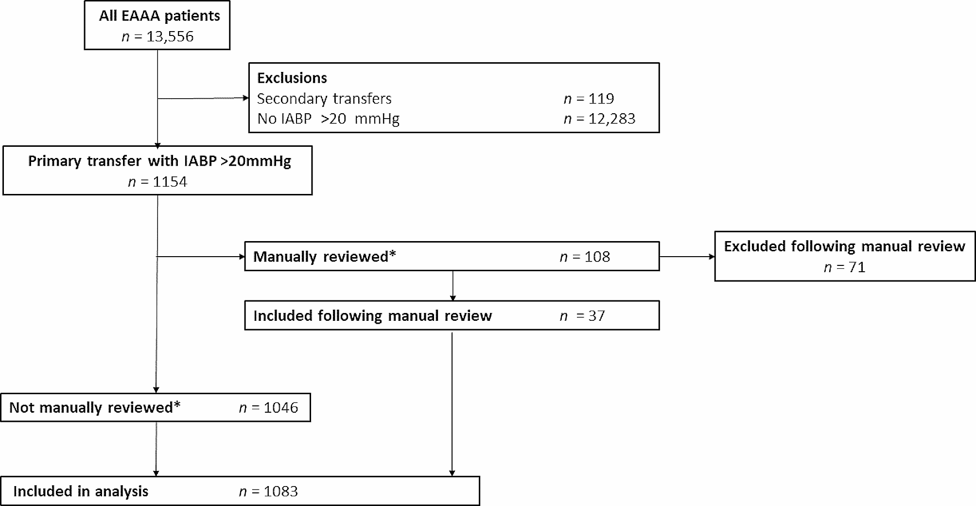
Fig. 1
Flow Chart of patients included in the study
Among the 29 women assessed by WBCT, two fetuses received a radiation dose above 100 mGy (110.89 mGy and 112.99 mGy, respectively). One of them (GA 18 weeks) was exposed to a radiation dose greater than 100 mGy in the context of repeated abdominal and pelvis imaging with and without arterial and venous phase contrast enhancement. For the second, fetal death was diagnosed before WBCT; concordantly, a higher dose was tolerated in the trauma appraisal of the mother.
Table 2 Patient’s characteristicsNine patients had a selective imaging strategy. In 3 of 54, the fetus was in the irradiation field, but the clinical assessment alone limited the injury appraisal to a selective strategy with regional CT and/or standard radiographs. This reduced the fetal radiation dose in one patient with multiple abdominal stab wounds to 51 mGy after a trunk-only scan. One patient had a standard pelvic X-ray, which was sufficient to rule out a pelvic fracture (fetal radiation dose < 1 mGy); one patient required two perioperative lumbar radiographs in the operating room after spine surgery (fetal radiation dose < 1 mGy). The initial assessment of this patient consisted of magnetic resonance imaging (MRI). For the 6 remaining patients, the fetus was not exposed to any radiation because it was out of the radiation field.
Table 3 Cumulated radiation dose (mGy) received by the fetus according to the type of injury assessment method and the gestational age Secondary outcomesMaternal mortalityThree pregnant women died as a consequence of their trauma (6%). One woman died on day one after multiple stab wounds to the trunk and initially resuscitated haemorrhagic cardiac arrest on scene. Two other patients succumbed after MVAs at 24 and 30 weeks gestation. One patient was in cardiac arrest on scene and died within a few hours of arrival due to uncontrolled haemorrhagic shock despite resuscitation and damage control laparotomy. The other patient suffered massive craniofacial trauma with severe intracranial hypertension due to brain oedema despite decompressive craniectomy. An emergency caesarean section was performed because of fetal bradycardia.
Fetal mortalityAn obstetric ultrasound was at admission performed to assess fetal vitality in 39 of 54 patients (72%), in the resuscitation room (90%) or in the first 24 h (10%). Five patients were considered too premature in their pregnancy to perform this exam. Nine of 54 fetuses (17%) died: one uterine rupture at 10 weeks, one early traumatic miscarriage following a high velocity MVA, six in utero fetal deaths and one fetus at 38 weeks requiring emergency caesarean section due to extreme bradycardia (maternal haemorrhagic shock) but was stillborn. Most of them (7 of 9) died upon arrival in the trauma bay as diagnosed by fetal ultrasound.
Two patients underwent therapeutic abortions after the discovery of polymalformative syndromes. The first was diagnosed in the context of trauma, and the second was diagnosed later in pregnancy following fetal irradiation of less than 1 mGy resulting from a clinically guided initial assessment.
Maternal haemorrhagic shockSix patients presented with haemorrhagic shock. Four patients required immediate surgery before WBCT. The two remaining patients underwent WBCT first after haemodynamic stabilization. In total, five patients required emergency surgeries: three splenic injuries, four hepatic injuries, one uterine rupture, and two wounds of the digestive tract (caecum and small intestine). One patient needed embolization of the two hypogastric arteries due to active haemorrhage complicating a pelvic fracture after an emergent caesarean section for fetal bradycardia on admission. Among those six patients, one died, while five fetuses did not survive.
Imaging injury assessmentAmong the 48 (89% of all patients) E-FAST exams performed upon arrival in the resuscitation room, 8 (16%) were positive: seven cases of haemoperitoneum, two of haemothorax and one of pneumothorax.
A WBCT was ordered for 42 of 54 patients (78%): 35 were performed immediately after the first clinical assessment in the resuscitation room, four after fetal extraction (two emergency caesarean sections with only one child alive and two intraoperative fetal extractions for deceased fetuses) and four after emergency surgery (exploratory laparotomy).
Nine patients (17%) had a clinically guided imaging strategy. These were haemodynamically stable, with a negative E-FAST and no clinical signs of severe injury. A patient who presented with a sensory-motor deficit of the lower limbs underwent lumbar spine MRI demonstrating a T12 burst fracture with spinal cord compression. Three had CT guided with clinical examination (head after gunshot, trunk after multiple stab wounds, head and spine after loss of consciousness and pain). Two patients had X-rays focused on painful areas (limbs, thorax). Three patients did not have any imaging other than E-FAST.
Among the 54 included patients, 9 of them were unknown and diagnosed during routine pregnancy testing on admission. Seven of them had a WBCT while 2 had clinically guided strategy. On the opposite, 7 fetuses died upon arrival in the trauma bay as diagnosed by fetal ultrasound, 6 of them having a WBCT. So if we try to calculate a WBCT rate where a viable pregnancy was known at the time of decision, this would be 29 WBCT with viable fetuses among 38 patients, so a WBCT rate of 76%.
留言 (0)