
記住我


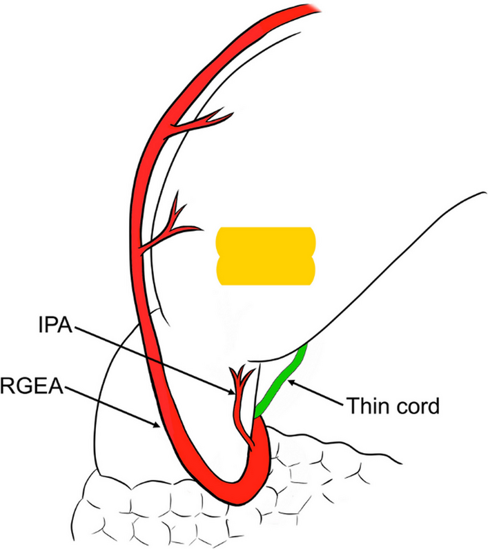
Here, we report a case of PPPV, which is an extremely rare congenital portal vein anomaly. Portal vein development begins at 4 weeks of gestation and is formed by the paired vitelline veins with three anastomoses that traverse the foregut (duodenum). As the weeks pass, the lower part of the right vitelline vein and the upper part of the left vitelline vein disappear, and the intermediate anastomosis on the dorsal side of the duodenum forms the portal vein main trunk [3, 4] (Fig. 6A, B). The development of the pancreas is established by fusion of the dorsal and ventral pancreatic buds arising from the foregut [5, 6]. The dorsal pancreatic bud arises ventrally to the left vitelline vein and the ventral pancreatic bud arises contralateral and slightly caudal to the dorsal pancreatic bud. As the duodenum rotates, the ventral pancreatic bud fuses behind the dorsal pancreatic bud, establishing the normal positional relationship between the portal vein and the pancreatic head (Fig. 6A, B). Regarding the etiology of PPPV, malposition of the dorsal pancreatic bud has been emphasized. Matsumoto et al. hypothesized that PPPV is established when the dorsal pancreatic bud arises on the dorsal side of the left vitelline vein [6]. Tomizawa et al. hypothesized that the formation of the dorsal pancreatic bud caudal to the intermediate anastomosis is the cause of PPPV [7] (Fig. 6C). We summarized 15 PPPV cases reported so far [6,7,8,9,10,11,12,13,14,15,16,17,18], including the present case (Table 1). Twelve of these 15 cases have been reported in Japan. In contrast to PDPV, which has been reported as a complication of other visceral malformations in childhood, all cases were adult cases, and only two of the reported cases of PPPV had congenital biliary dilatation, and most of them were reported to have PPPV during examination and treatment for other diseases. In addition to the abnormality in which the portal vein runs in front of the pancreas and behind the duodenum, this disease has the following characteristic findings. The portal vein was L-shaped or inverted L-shaped in ten cases, and in 11 cases, it passed through the ventral or right side of the common bile duct. Five cases with abnormal branching of the portal vein were also reported, most of which involved early branching of the portal vein. Moreover, of the 13 PPPV cases for which information on portal vein morphology was provided, seven (53.8%) had irregularly dilated or winding portal vein in the hepatic hilum, and there were five cases with a fragile and thin portal vein wall and firm adhesion with the surrounding tissue. Five of these patients underwent pancreaticoduodenectomy and three underwent combined resection of the portal vein because of difficulty in dissecting the portal vein [9, 16, 18], and two of them had intraoperatively massive bleeding while isolating the portal vein, and thrombosis in the reconstructed portal vein [16, 18]. Shimizu et al. reported the difficulty of portal vein reconstruction due to the thinness of the portal vein wall and the difficulty of postoperative management due to complications of portal vein thrombosis [16].
Fig. 6
Development of the portal vein. A Paired vitelline veins are connected by three anastomoses. The duodenum lies ventral to the intermediate anastomosis. The dorsal pancreatic bud arises ventrally to the left vitelline vein, and the ventral pancreatic bud arises contralateral and slightly caudal to the dorsal pancreatic bud. B Normal development of portal vein and pancreas. The lower part of the right vitelline vein and the upper part of the left vitelline vein disappear, and the intermediate anastomosis on the dorsal side of the duodenum forms the portal vein main trunk. As the duodenum rotates, the ventral pancreatic bud fuses behind the dorsal pancreatic bud. C Development of the prepancreatic postduodenal portal vein. The dorsal pancreatic bud arises on the dorsal side of the left vitelline vein, or arises caudal to the intermediate anastomosis, which results in the portal vein posterior to the duodenum but anterior to the pancreas. SV sinus venosus, d dorsal pancreatic bud, v ventral pancreatic bud, VV vitelline vein, UV umbilical vein, PV portal vein, PPPV prepancreatic postduodenal portal vein
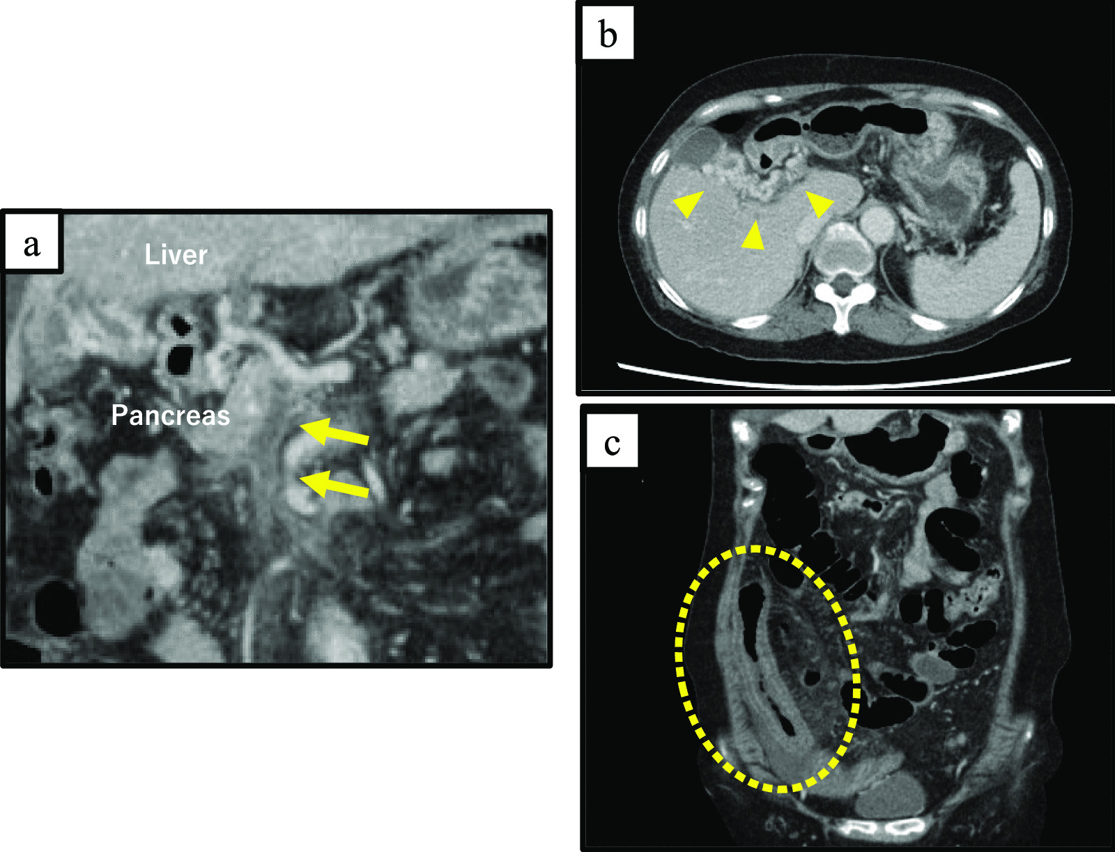
Table 1 Previous reports of prepancreatic postduodenal portal veinIn our case, PPPV and the complex morphology of the portal vein could be identified by evaluating the contrast-enhanced CT and 3D-constructed images as preoperative examinations for HCC. His portal vein had an inverted L-shaped formation and ran along the ventral side of the bile duct, which is consistent with previous reports. In addition, the portal vein was dilated at the hepatic hilum, complicatedly meandering, bending, and branching, similar to cavernous transformation, with some porto-portal communications observed. Among the previously reported PPPV cases, the complex morphology of this case is extremely rare. We chose S8 partial resection as the operative procedure instead of anatomical resection to avoid manipulation around the hepatic hilum because of the anomaly of the portal vein and the location of the HCC on the liver surface. However, anatomical liver resection using the staining method without touching the hepatic hilum was an option to consider. At the time of resection, we considered the possibility that his portal vein wall was thinner and more fragile than usual, as pointed out by previous reports, and did not perform the Pringle maneuver, even though there was no scientific evidence that this technique was dangerous for PPPV. Fortunately, no complex meandering or irregular dilation was observed in the peripheral portal vein branches. Therefore, a partial hepatectomy for the tumor on the surface of the liver could be performed without affecting the malformation of the portal vein. If the tumor was located near the hepatic hilum, the risk of resection was thought to be markedly increased.
留言 (0)