Protein dynamics by the combination of high-speed AFM and computational modeling
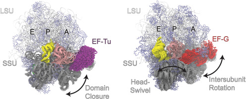
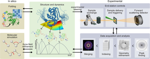
Understanding how proteins function, a major goal in biophysics, can be facilitated by the direct observation of individual single molecules during their functional activity. It is only high-speed atomic force microscopy (HS-AFM) that can perform this direct observation of single molecules [1]. Thus far, more than 200 different proteins have been filmed with HS-AFM at ∼100 ms temporal, 2–3 nm lateral, and ∼0.1 nm vertical resolution [2∗,3], e.g., myosin V walking on actin filaments [4] and the stator ring of F1-ATPase with conformational and chemical states propagating over the subunits in one direction [5]. The time resolution is now approaching 10 ms by the developments of a new scanning mode [6], a faster Z-scanner [7], and other underlying techniques, which can significantly expand the range of observable protein systems and dynamic phenomena. However, there is an intrinsic limitation in AFM imaging; only the surface topography can be acquired, and the AFM tip is too large to resolve details on the sub-nanometer scale. Therefore, the interpretation of the captured images is often challenging, and an atomistic-level understanding of the functional mechanism seems impossible. In this situation, post-experimental analysis employing computational methods plays an increasingly important role. It can complement the experimental observations and facilitate the interpretation and understanding of HS-AFM images. Here we shall review recent developments and the status quo in this theoretical field. The current approaches can be distinguished into two main categories. On one side, modeling and simulation methods are developed to construct 3D conformations with atomistic resolution from topographic AFM images with limited resolution. On the other side, analysis methods are developed and applied to improve quantitative analysis of measured topographies and to compute their higher-resolution maps.


留言 (0)