
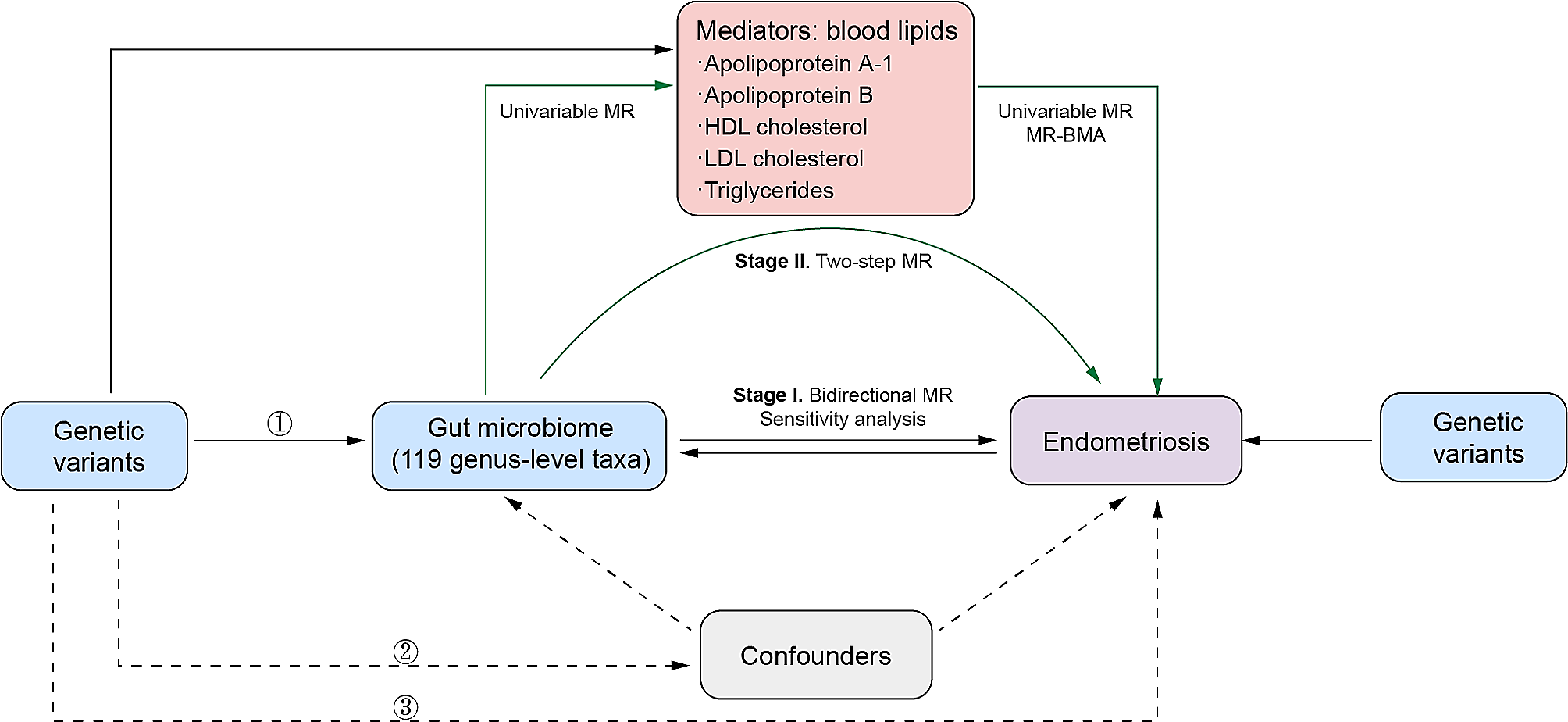
記住我


To clarify that AS appeared in model mice, The aortic arch of three groups of mice was stained with oil red O staining. The results found that the aortic arches of the C57BL/6 mice had no plaque, while the aortic arches of the ApoE−/− + NF group mice and ApoE−/− + HF group mice had obvious lipids deposition (Fig. 1A-B). Next, the researchers observed the presence of lipids to clarify the lipid status of mice in each group, Four items of blood lipids in collected mouse plasma were detected by kits. The results represented that by comparison with the C57BL/6 mice, the levels of four items of blood lipids in two groups of ApoE−/− mice were significantly upgraded, and the HF diet worsened the plasma lipid of ApoE−/− mice (Fig. 1D, left). Since the ratio of LDL-c/HDL-c is positively related to the pressure of cholesterol transportation, the researchers have made statistics on the above indicators of the three groups of mice and found that two ApoE−/− groups mice have significant plasma cholesterol transport loads which were heavier than that of mice in the C57 group (Fig. 1D, right). To explore which specific components of ApoE−/− mice’s blood lipids can be affected by HF, non-targeted lipidomics research has been used to further clarify the lipid categories that produce significant changes. According to the types of lipid metabolites, the results showed that the four main lipid components of CE, TAG, FFA, and PL in the mouse plasma were significantly up-regulated after giving HF diet, (Fig. 1E). The volcano map made of the detected lipid metabolites intuitively shows that there are significant changes in the plasma of the two groups of ApoE−/− mice, and it can be seen that there are more types of lipid metabolites that are significantly up-regulated than down-regulated metabolites (Fig. 1F). To explore the differences in the plasma as a whole, the researchers analyzed the detected plasma metabolites by PCA and PLS-DA. The similarity of lipid metabolites in each group can be expressed by the distance between samples. The farther the distance, the greater the difference between samples, which is reflected in the types and relative abundance of lipids contained. The clustering results of the two mathematical models intuitively show that the HF diet can produce obvious differences in plasma lipid metabolites of ApoE−/− mice (Fig. 1G-H). Finally, the researchers have made statistics on the lipid components detected in the plasma of the mice in this research. The heat map and the box plot showed that compared with the mice in the ApoE−/− + NF group, 10 types of CE, 20 types of TAG, 30 types of PL, 2 types of FFA, 2 types of sphingomyelin (SM), 1 types of acylcarnitine (Acy), and 1 type of ceramide (Cer) were significantly transformed in the plasma of the ApoE−/− + HF group (Fig. 1I-M). These results confirmed that the continuous high-cholesterol diet leads to abnormal blood lipid metabolism, and provided a basis for us to emphasize the intestinal intake of cholesterol.
Fig. 1
Atherosclerotic lesions and abnormal blood lipid levels of ApoE−/− mice fed a high-cholesterol diet. A-B Representative images of oil red O staining of the aortic arch and quantitative analysis of the percentage of oil red O positive staining area. Original magnification: 40 × . C Representative transmission electron microscope images of subcutaneous lipid droplets in the aortic arch. Original magnification: 1.2 k × . D The four levels of blood lipids (left) and the ratio of low-density lipoprotein to high-density lipoprotein (right) by kits. E Lipids in plasma samples were extracted and detected by HPLC-Q-TOF/MS. F Volcano diagrams of the ApoE−/− + NF group and the ApoE−/− + HF group. G PCA score chart, showing the difference between the C57BL/6 + NF group (green), the ApoE−/− + NF group (blue), and the ApoE−/− + HF group (red) in plasma samples. H PLS-DA score chart, showing the difference between the C57BL/6 + NF group (green), the ApoE−/− + NF group (blue), and the ApoE−/− + HF group (red) in plasma samples. I The heat map showed the difference in plasma lipid levels in each group. J-M The box plot reflected the relative content of each type of lipid. In all experiments, n = 6, the P value indicates the comparison with the ApoE−/− + NF group. Values are expressed as mean ± SEM
High-cholesterol diet-induced jejunum tissue lesions and abnormal jejunum lipid metabolism in ApoE−/− miceTo clarify the influence of a high-cholesterol diet aiming at jejunum lipid intake, the researchers first observed the morphology and structure of the jejunum of the three groups of mice by HE staining. The results showed the length of the jejunum villi of the mice in the ApoE−/− + HF group was significantly shorter than two groups of mice fed the NF diet, swelling, the depth of intestinal crypts increases significantly, showing a higher level of pathology (Fig. 2A-B). Oil red O staining suggested that the volume and number of lipid droplets in jejunum villi in the ApoE−/− + HF group was significantly larger than that in the C57BL/6 + NF group and ApoE−/− + NF group. (Fig. 2C-D). Similar to our study in plasma, to clarify the types of lipids accumulated in the jejunum, the researchers subsequently conducted a non-targeted lipidomics study on the jejunum of the mice in this research. According to the types of lipid metabolites, the total content of the four types of lipid components in the jejunum was counted. The researchers found that the three main lipid components CE, TAG, and FFA in the jejunum of mice in the ApoE−/− + HF group were significantly increased than those in ApoE−/− + NF (Fig. 2E). The volcano map made of the detected lipid metabolites intuitively shows that there are significant changes in the jejunum of the two groups of ApoE−/− mice, and it can be seen that there are more types of lipid metabolites that are significantly up-regulated than down-regulated metabolites (Fig. 2F). PCA and PLS-DA were used to analyze the detected metabolites of jejunum, to explore the differences between groups of jejunum as a whole. The clustering results of the two mathematical models visually suggested the ApoE−/− + NF group and the ApoE−/− + HF group had a clear difference in jejunum lipid metabolites (Fig. 2G-H). Finally, the results of the heat map and the box plot suggested that compared with the ApoE−/− + NF group, 6 types of CE, 27 types of TAG, 15 types of FFA, 3 types of PL, and 2 types of SM in the jejunum of mice in the ApoE−/− + HF group had significant change (Fig. 2I-M). The researchers found that CE containing 16:1, 18:1, and 20:3 ester acyl groups increased synchronously and significantly in both jejunum and plasma. Later, we detected the TC and free cholesterol (FC) content in the contents of the distal colorectal and proximal small intestine of mice by kits, and the ratio can reflect the ability of the small intestine to absorb cholesterol. We found that the absorption fraction of exogenous cholesterol in mice fed the NF diet was about 50%, while that in mice fed the HF diet was about 35% (Fig. 2N-O). Such results suggested that under the condition of a high-cholesterol diet, a large amount of cholesterol is absorbed into the blood by the intestine.
Fig. 2
Intestinal lesions and abnormal intestinal lipid levels in ApoE−/− mice fed a high-cholesterol diet. A-B Representative images of HE staining of the small intestine and evaluation scores of intestinal villi morphology. C-D Representative images of oil red O staining of proximal jejunum villi and quantitative analysis of the percentage of oil red O positive staining area. Original magnification: 40 × . E The lipids of small intestine samples were extracted, detected by HPLC-Q-TOF/MS, and statistically analyzed with different mathematical models. F Volcano diagrams of the ApoE−/− + NF group and the ApoE−/− + HF group. G PCA score chart, showing the difference between the C57BL/6 + NF group (green), the ApoE−/− + NF group (blue), and the ApoE−/− + HF group (red) in the small intestine samples. H The PLS-DA score chart shows the difference between the C57BL/6 + NF group (green), the ApoE−/− + NF group (blue), and the ApoE−/− + HF group (red) in the small intestine samples. I The heat map shows the difference in the small intestine lipids in each group. J-M The box plot reflects the relative content of each type of lipid. N–O Total cholesterol and free cholesterol in intestinal contents tested by kits. In all experiments, n = 5, the P value indicates the comparison with the ApoE−/− + NF group. Values are expressed as mean ± SEM
High-cholesterol diet significantly activated the expression of LXRα in the jejunum of ApoE−/− miceThe jejunum can not only transport exogenous lipids into the blood in different ways but also can synthesize lipids de novo. To clarify that a high-cholesterol diet can cause the response of key indicators of lipid metabolism in the jejunum, RT-PCR, WB, IHC, and IF were used to study the expression of key lipid metabolism indexes in the jejunum of three groups of mice. The results showed that the high-cholesterol diet can significantly activate the jejunum mRNA levels of NPC1L1, ABCG5, ABCG8, ABCA1, and LXRα in the ApoE−/− + HF mice, but had no statistical change in the ACC, ACS, SREBP1, HMGCR, and LXRβ (Fig. 3A). Then we analyzed the expressions of NPC1L1, ABCG5, ABCG5, ABCA1, and LXRα in the jejunum and liver by WB. We found that a normal diet could not activate the above targets in the jejunum, even if ApoE was knocked out. A high-fat diet can significantly up-regulate the protein expression of the above targets. Different from jejunum, the above targets in the liver were significantly up-regulated when ApoE was knocked out, and a high-fat diet could further up-regulate the protein expression. These results suggested that jejunum lipid metabolism was more biased towards the activation of exogenous lipids, while the liver needs to take into account the metabolism of endogenous lipids and exogenous lipids, and is sensitive to any factors that lead to lipid metabolism. Then the IF, and IHC results showed that compared with the ApoE−/− + NF group, the protein expression of NPC1L1, ABCG5, ABCG8, and LXRα in the ApoE−/− + HF group mice was significantly increased (Fig. 3E-J). These results indicated that a high-cholesterol diet can activate intestinal epithelial cells to excrete exogenous cholesterol mediated by LXRα and ABCG5/G8. Therefore, it can reduce the negative influence of lipid uptake in the jejunum due to the activation of NPC1L1 to a certain degree. However, the regulation mechanism of cholesterol uptake cannot reverse the transport of exogenous cholesterol into the blood by the jejunum.
Fig. 3
The influence of a high-fat diet on key targets of jejunum lipid metabolism in ApoE−/− mice. A The mRNA expression of targets related to lipid metabolism in the jejunum. B-D Representative western blots and relative quantitative analysis of NPC1L1, ABCG5, ABCG8, and ABCA1 in the proximal jejunum and liver. E Representative IHC staining images and quantitative analysis of LXRα and NPC1L1 in the jejunum. F-K Representative IF images and quantitative analysis of ABCG5 and ABCG8 in the proximal jejunum. In all experiments, n = 5, the P value indicates the comparison with the ApoE−/− + NF group. Values are expressed as mean ± SEM
The efflux of excessive intracellular cholesterol by Caco-2 cells is mediated by the LXRα-ABCG5/G8 pathwayUsing Caco-2 cells, the researchers studied the influence of high cholesterol culture on lipid metabolism. The Caco-2 cells were cultured at the bottom of the culture dish, and then the micelles with the final concentration of 100 μM cholesterol were added to the culture medium. By examining the IF results of tight junction protein ZO-1, we found that the protein level of ZO-1 cultured with high cholesterol was significantly lower than that in normal cells (Fig. 4A-B). By observing the size and number of lipid droplets in the oil red O stained image, the researchers found the accumulation of intracellular lipids in Caco-2 cells in the Model group (Fig. 4C-D). Subsequently, through the different lipids test kits, researchers can evaluate the relative content of different types of lipid components in cells. These results suggested that the contents of various lipids in Caco-2 cells increased significantly after the induction of high cholesterol (Fig. 4E-H). To clarify the targets that have significantly changed in Caco-2 cells, the researchers first analyzed the mRNA expression of key lipid metabolism targets by RT-PCR. The above results suggested that the mRNA levels of LXRα, NPC1L1, ABCG5, and ABCG8 were increased significantly (Fig. 4I). The IF results for the above targets were consistent with the RT-PCR results (Fig. 4J-K). Next, the researchers studied the changes in cholesterol metabolism in Caco-2 cells after LXRα was inhibited by giving GSK2033, a specific inhibitor of LXRα and siRNA targeting Continuous application of GSK2033 at 10, 20, and 40 nM or LXRα siRNA at 25, 50, and 100 nM for 24 h can further lead to intracellular lipid accumulation. Moreover, the application of 20 nM GSK2033 or 50 nM LXRα siRNA for 12, 24, and 48 h can lead to the aggravation of intracellular lipid accumulation (Fig. 4L-P). Severe lipid accumulation can be observed in Caco-2 cells by applying 20 nM GSK2033 or 50 nM LXRα siRNA 24 h in advance, the researchers tried to explore the expression of NPC1L1, ABCA1, ABCG5, and ABCG8 when LXRα was inhibited. The researchers found that the protein levels of ABCA1, ABCG5, and ABCG8 were significantly reduced but the expression of NPC1L1 did not change significantly (Fig. 4Q-R).
Fig. 4
The cholesterol metabolism of intestinal Caco-2 cells is mediated by LXRα. A-B Representative IF images and quantitative analysis of ZO-1 in Caco-2cells. C-D Representative images of oil red O staining of Caco-2 cells and quantitative analysis of the percentage of oil red O positive staining area. E–H Lipid levels in Caco-2 cells tested by kits. I The mRNA expression of targets related to lipid metabolism in Caco-2 cells. J-K Representative IF images and quantitative analysis of NPC1L1, ABCG5, ABCG8, LXRα in Caco-2cells. L-P representative images of oil red O staining of Caco-2 cells incubated with 100 μmol of cholesterol for 24 h and quantitative analysis of the positive area; Q-R Representative Western blot and relative quantitative analysis of NPC1L1, ABCA1, ABCG5, ABCG8, and LXRα after incubating Caco-2 cells with 100 μmol of cholesterol for 24 h. In the above experiments, n = 5, the P value indicates the comparison with the control group. Values are expressed as mean ± SEM
留言 (0)